Infrastructure and technology development
CCFS links three internationally recognised concentrations of analytical geochemistry infrastructure: GEMOC’s Geochemical Analysis Unit (Macquarie University) and the associated Computing Cluster, the Centre for Microscopy, Characterisation and Analysis (UWA/Curtin) and the John de Laeter Centre of Mass Spectrometry. All are nodes for the NCRIS AuScope and Characterisation Capabilities, and have complementary instrumentation and laboratories. In addition, Curtin and UWA share a leading facility for paleomagnetic studies, and facilities for experimental mineralogy and petrology are being built up at Macquarie and Curtin.
Facilities:
CCFS/GEMOC INFRASTRUCTURE, LABORATORIES AND INSTRUMENTATION
CMCA TECHNOLOGY DEVELOPMENT AND INSTRUMENTATION
WESTERN AUSTRALIA PALAEOMAGNETIC AND ROCK-MAGNETIC FACILITY
CCFS/GEMOC INFRASTRUCTURE, LABORATORIES AND INSTRUMENTATION
The analytical instrumentation and support facilities of the Macquarie University Geochemical Analysis Unit (GAU) represent a state-of-the-art geochemical facility.
The GAU contains:
• a Cameca SX-100 electron microprobe
• a Zeiss EVO MA15 Scanning electron microscope (with Oxford Instruments Aztec Synergy EDS/EBSD)
• four Agilent quadrupole ICPMS (industry collaboration; two 7500cs; two 7700cx)
• a Nu Plasma multi-collector ICPMS
• a Nu Plasma high resolution multi-collector ICPMS
• a Nu Attom high resolution single-collector sector field ICPMS
• a Thermo Finnigan Triton TIMS
• a custom-built UV laser microprobe, usable on the Agilent ICPMS
• three New Wave laser microprobes (one 266 nm, two 213 nm, each fitted with large format sample cells) for the MC-ICPMS and ICPMS laboratories (industry collaboration)
• a Photon Machines Excite Excimer laser ablation system
• a Photon Analyte G2 Excimer laser ablation system
• a Photon Machines Analyte198 Femto-second laser ablation system
• a PANalytical Axios 1kW XRF with rocker-furnace sample preparation equipment
• a LECO RC412 H2O-CO2 analyser
• an Ortec Alpha Particle counter
• a New Wave MicroMill micro-sampling apparatus
• a ThermoFisher iN10 FTIR microscope
• selFrag electrostatic rock disaggregation facility
• clean labs and sampling facilities provide infrastructure for ICPMS, XRF and isotopic analyses of small and/or low-level samples
• experimental petrology laboratories include four piston-cylinder presses (pressures to 4 GPa), hydrothermal apparatus, controlled atmosphere furnaces, Griggs apparatus and a multi-anvil apparatus for pressures to 27 GPa
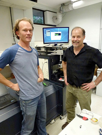
Will Powell and Jamie Barbula, (Photon Machines) during the installation of the Femto-second laser.
THE GEMOC FACILITY FOR INTEGRATED MICROANALYSIS (FIM) AND MICRO-GIS DEVELOPMENT
GEMOC is continuing to develop a unique, world-class geochemical facility, based on in situ imaging and microanalysis of trace elements and isotopic ratios in minerals, rocks and fluids. The Facility for Integrated Microanalysis now consists of four different types of analytical instrument, linked by a single sample positioning and referencing system to combine spot analysis with images of spatial variations in composition (“micro-GIS”). All instruments in the FIM have been operating since mid-1999. Major instruments were replaced or upgraded in 2002-2004 through the $5.125 million DEST Infrastructure grant awarded to Macquarie University with the Universities of Newcastle, Sydney, Western Sydney and Wollongong as partners. In late 2009 GEMOC was awarded an ARC LIEF grant to integrate the two existing multi-collector inductively-coupled-plasma mass spectrometers (MC-ICPMS) with 3 new instruments: a femtosecond laser-ablation microprobe (LAM); a high-sensitivity magnetic-sector ICPMS; a quadrupole ICPMS. The quadrupole ICP-MS was purchased and installed in 2010; a Photon Machines femtosecond laser system was installed in June 2012; and a Nu Attom ICP-MS was installed in January 2013. In 2012 GEMOC was awarded ARC LIEF funding for a second generation MC-ICPMS. Evaluation of the current instruments on the market was undertaken in 2013 and an order will be placed in early 2014.
EQUIPMENT FOR HIGH-PRESSURE EXPERIMENTATION
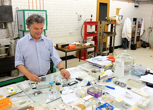
Expansion of the facilities for high-pressure experimentation at GEMOC began in 2013. The laboratories now house two piston-cylinder presses (pressures to 3 GPa), a Griggs apparatus and a multi-anvil apparatus for (pressures to 27 GPa). Funding for the purchase of a diamond-anvil apparatus for use at much higher pressures, and an additional large-volume multi-anvil apparatus were acquired in 2013, expanding the experimental capability for mantle studies.
Raymond Jones visited Simon Clark in March - April -2013 to help set up the high pressure lab.
PROGRESS IN 2013:
1. Facility for Integrated Microanalysis
a. Electron Microprobe: The Zeiss EVO MA15 SEM carried the electron imaging workload providing high-resolution BSE and CL images for TerraneChron® (http://www.gemoc.mq.edu.au/TerraneChron.html) and all other research projects, including diamonds and diamondites, PGM in chromitites and experimental petrology. The Oxford Instruments AZtec Synergy combined Energy Dispersive System and Electron Back-Scatter Diffraction detector installed in 2012 has enabled simultaneous elemental and crystal orientation mapping. This capability has enabled new research directions in the study of deformation processes in mantle and crustal rocks, including melt/rock interaction in high-grade metamorphic rocks and metasomatism of mantle-derived peridotites, pyroxenites and chromitites. The Cameca SX100 electron microprobe continued to service the demands for quantitative mineral analyses and X-ray composition maps for all projects including analysis of chromites; analysis of base metal sulfides and platinum group minerals; minor and trace element analysis of metals. A Bruker Energy Dispersive Spectrometer system was installed on the SX100 and was purchased with funding provided by a MQSIS 2013 Faculty of Science Infrastructure Fund grant.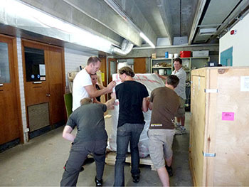
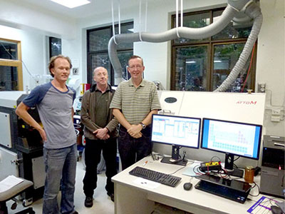
Will Powell, Mark Chandler (Nu Instruments) and Norman Pearson (Photon Machines) oversee the installation of the Nu Attom.
b. Laser-ablation ICPMS microprobe (LAM): In 2013 the LAM laboratory was used by 12 Macquarie PhD thesis projects, eleven international visitors, two Honours students, nine users from other Australian institutions and several in-house funded research projects and industry collaborations. Projects included the analysis of trace elements in the minerals of mantle-derived peridotites, pyroxenites and chromitites, in sulfide minerals and in a range of unusual matrices. U-Pb analysis of zircons was again a major activity with geochronology projects (including TerraneChron® applications: http://www.gemoc.mq.edu.au/TerraneChron.html) from Australia (NSW, WA, SA), New Zealand, Algeria, Australia, Bostwana, Brazil, Indonesia, Laos, PNG and Russia. Method development was also undertaken for the U-Pb dating of baddeleyite and rutile. The LAM laboratory also routinely provides data for projects related to mineral exploration (diamonds, base metals, Au) as a value-added service to the industry.
c. MC-ICPMS: The high demand for LAM MC-ICPMS time for in-situ high-precision ratio measurements was again led by the analysis of Lu-Hf isotopes in zircon as a major strand of the TerraneChron® activities (see http://www.gemoc.mq.edu.au/TerraneChron.html) and Re-Os dating of single grains of Fe-Ni sulfides and alloys in mantle-derived rocks. In situ Hf isotopes were measured in zircons from Australia (NSW, WA, SA), New Zealand, Algeria, Australia, Bostwana, Brazil, Indonesia, Laos, PNG and Russia. Re-Os studies were undertaken on xenoliths from eastern China, Siberia, Mongolia, Italy, Spain and South Africa, and sulfide and platinum group minerals in chromitites from Cuba, Spain, Turkey.
d. Laboratory development: The clean-room facility established in 2004 continued to be used primarily for isotope separations for analysis on the Triton TIMS and Nu Plasma MC-ICPMS. Routine procedures have been established for Rb-Sr, Nd-Sm, Lu-Hf and Pb isotopes, as well as U-series methods (U, Th and Ra). Further refinement was undertaken of methods for whole-rock Re-Os isotopes of basaltic rocks and the adaptation of conventional techniques for Rb-Sr and Nd-Sm isotope separation to the nano-scale to process small sample sizes.
Further development of ‘non-traditional’ stable isotopes was carried out in 2013 and focused on Mg isotopes in mantle minerals and for the separation and analysis of Li isotopes in granitic rocks and in clinopyroxene from mantle-derived eclogites and peridotites.
e. Software: GLITTER (GEMOC Laser ICPMS Total Trace Element Reduction) software is our on-line interactive program for quantitative trace element and isotopic analysis and features dynamically linked graphics and analysis tables. This package provides real-time interactive data reduction for LAM-ICPMS analysis, allowing inspection and evaluation of each result before the next analysis spot is chosen. GLITTER’s capabilities include the on-line reduction of U-Pb data. Sales of GLITTER are handled by Access MQ and GEMOC provides customer service and technical backup. During 2013 a further 11 full licences of GLITTER were sold bringing the total number in use to more than 220 worldwide, predominantly in earth sciences applications but with growing usage in forensics and materials science. Dr Will Powell continued in his role in GLITTER technical support and software development through 2013. The current GLITTER release is version 4.4.4 and is currently available without charge to existing customers and accompanies all new orders.
 2. X-Ray Fluorescence Analysis
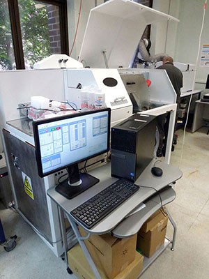
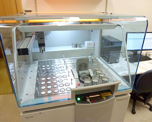
In November 2012 a PANalytical Axios 1 kW X-ray Fluorescence Spectrometer was installed (see photo below ). This instrument is a wavelength spectrometer system and replaced the Spectro XLAB2000 energy-dispersive X-ray spectrometer, which was installed in November 2000. The Axios instrument is used routinely to measure whole-rock major element compositions on fused glass discs and trace-element concentrations on pressed-powder pellets. In 2013 the sample preparation equipment was upgraded and included a new furnace to make high-quality cast glass beads.
3. Whole-rock solution analysis
An Agilent 7500cs ICPMS produces trace-element analyses of dissolved rock samples for the projects of CCFS/GEMOC researchers and students and external users, supplementing the data from the XRF.
The ICPMS dedicated to solution analysis is also used to support the development of ‘non-traditional’ stable isotopes with the refinement of separation techniques and analytical protocols (see 1. d above ).
4. Diamond preparation and analysis
The GEMOC laser-cutting system (donated by Argyle diamonds in 2008) was used during 2013 to cut thin plates of single diamond crystals as part of the on-going research into diamond genesis. The plates are used for detailed spatial analysis of trace elements, isotopic ratios and the abundance and aggregation state of nitrogen. The nitrogen measurements are made using the ThermoFisher iN10FTIR microscope, which allows the spatial mapping of whole diamond plates at high resolution with very short acquisition times.
5. selFrag – a new approach to sample preparation
GEMOC’s selFrag instrument was installed in May 2010 and was the first unit in Australia. This instrument uses high-powered electrical pulses to disaggregate rocks and other materials along the grain boundaries. It removes the need to crush rocks for mineral separation, and provides a higher proportion of unbroken grains of trace minerals such as zircon. Since its installation, selFrag usage has continued to grow and in 2013 it was used for a range of applications including zircon separation, the analysis of grain size and shape in complex rocks, and the liberation of trace minerals from a range of mantle-derived and crustal rocks. Method development also continued in 2013 on the CNT Hydroseparator for the separation of small volumes of ultrafine material (e.g. alloys in chromitites).
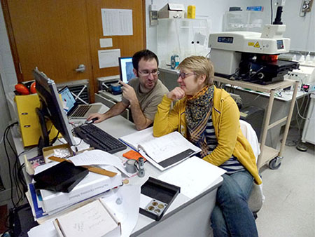
José María González Jiménez and Irina Tretiakova working on the LA-ICPMS.
6. Computer cluster
The cluster Enki has continued to be a workhorse for the geodynamics group, supporting two funded research projects, 3 PhD projects, a postdoc, and numerous masters-level projects. Recent developments have included the migration of the new mantle convection code Aspect (based on the deal.II finite element libraries) to Enki, and continued development of this code, led by Siqi Zhang, has led to a number of capability breakthroughs, including coupled core-mantle simulations, integrated mantle melting/convection simulations, and coupled plate/fluid flow models.
CMCA TECHNOLOGY DEVELOPMENT AND INSTRUMENTATION
The University of Western Australia’s Centre for Microscopy, Characterisation and Analysis (CMCA) is a $40M core facility providing analytical solutions across a diverse array of scientific research. The world-class facilities and associated technical and academic expertise are the focus of micro-analytical and characterisation activities within Western Australia, while strong links and collaborations have earned the CMCA an excellent national and international reputation. The CMCA incorporates the Western Australian Centre for Microscopy, and is a node of the NCRIS Characterisation capabilities, the National Imaging Facility (NIF) and the Australian Microscopy and Microanalysis Research Facility (AMMRF). It is also associated with the NCRIS funded Australian National Fabrication Facility (ANFF), and AuScope, which have made a substantial contribution to facilities run by CMCA.
CMCA capabilities:
• CAMECA IMS 1280 and CAMECA NanoSIMS 50 ion microprobes
• JEOL JXA 8530F electron microprobe, with Cathodoluminescence imaging (CL)
• Transmission Electron Microscopes (JEOL 2100 and 2000FX), with EDS and energy-filtered EELS analysis capabilities
• Scanning electron microscopes (Zeiss 1555, 2 JEOL 6400, Philips XL30)
• X-ray powder diffraction (Panalytical Empyrean)
• NMR spectroscopy (2 Bruker Avance and 2 Varian spectrometers)
• Optical and confocal microscopy
• Micro-CT (Skyscan 1176)
• Bioimaging, flow cytometry, cell sorting, and laser micro-dissection
• X-ray crystallography
• GC and HPLC mass spectrometry
• Biological sample cryo-preparation and ultramicrotomy
THE AMMRF FLAGSHIP ION PROBE FACILITY
The CAMECA IMS 1280 and NanoSIMS 50 are flagship instruments of the Australian Microscopy & Microanalysis Research Facility (AMMRF). The AMMRF Flagship Ion Probe Facility offers state-of-the-art secondary ion mass spectrometry (SIMS) capabilities to the Australian and international research communities, allowing in-situ, high-precision isotopic and elemental analyses, secondary ion imaging, and depth profiling on a wide range of samples.
The IMS 1280 large-geometry ion probe, installed in 2009, was co-funded by the University, the State Government of Western Australia, and the Federal Government’s Department of Innovation, Industry, Science and Research (DIISR) under the ‘Characterisation’ (AMMRF) and ‘Structure and Evolution of the Australian Continent’ (AuScope) capabilities of the National Collaborative Research Infrastructure Strategy (NCRIS). The NanoSIMS 50, installed in 2003, was funded through the Federal Government’s NCRIS-precursor, the Major National Research Facility scheme (NANO-MNRF).
The instruments are managed by the Western Australia Ion Probe Management Committee, which also manages the two SHRIMP II ion probes located at Curtin University. Access to the Ion Probe Facility is subsidised for publicly-funded researchers within Australia via a merit-based competitive application scheme, where projects are assessed by a scientific committee of international experts.
The Ion Probe Facility is a key characterisation component within the ARC Centre of Excellence for Core to Crust Fluid Systems. To ensure the highest levels of quality and throughput, the CCFS has provided funding for a Research Associate position within the Ion Probe Facility, to facilitate direct scientific and technical interaction for all CCFS users and projects.
PROGRESS IN 2013:
Recent successes in ARC LIEF funding will update the electron microscopy facilities, with the addition of a FEI Titan TEM and a FEI Verios 460 SEM. The CMCA will also receive funding for a new Cameca NanoSIMS 50L ion probe through CSIRO’s Science and Industry Endowment Fund (SIEF). The new instrument will be part of the tripartite Advanced Resources Characterisation Facility, along with a Geo-Atom Probe to be located at Curtin University, and the in-house development of a MAIA mapping facility at CSIRO. The NanoSIMS 50L represents a considerable technological advance over the existing NanoSIMS, with a seven-FC/EM multicollector array and a new oxygen ion source allowing high-resolution isotope measurements on geological samples.
In 2013, the Ion Probe Facility has continued to contribute to various projects in the context of CCFS. These have included a wide range of topics, from the characterisation of mantle metasomatism (O isotopes in garnet, J Huang), the origin of diamonds (C isotopes in diamond: W Griffin and Z Spetsius), magmatic processes and crustal growth (O isotopes in zircon; E Belousova), the origin of ore deposits (S isotopes in sulfides; M Fiorentini). In addition, the NanoSIMS has also been involved in the measurement of element diffusion across mineral interfaces, trace element transportation along grain boundaries, and S isotopes (D. Wacey, M. Fiorentini).
High-precision isotope measurement with SIMS requires calibration against known standards to correct for instrumental mass fractionation between analysis sessions. This varies significantly between different materials, such that each new material analysed by SIMS necessitates the development of new standards. Standards are in constant development at CMCA and currently include pyroxene and olivine (O isotopes) as well as a range of Si-bearing materials (SiC, Si metal, silicates) for Si isotopes. Development continues on diamond (C isotopes), lawsonite, pyroxene, garnet and olivine (O isotopes), tourmaline and serpentine (B isotopes), pentlandite, pyrrhotite and chalcopyrite (S and Fe isotopes). The development of standards for unknown isotope systems aims to identify potentially new geochemical tools.
Personnel:
2013 saw a number of staff changes at the CMCA. Laure Martin and John Cliff continued to develop standards and analytical protocols for isotopic measurement using the Cameca IMS 1280 ion probe. Matt Kilburn spent six months on sabbatical at Harvard University, learning about the intricacies of applying NanoSIMS to biomedical research. David Wacey returned from overseas to work full time on CCFS Foundation Project 5. Rong Liu left the CMCA to take up a position running the SIMS lab at the University of Western Sydney. His position was filled by Paul Guagliardo. David Adams left CMCA to take up a similar position as EPMA operator at GEMOC, Macquarie University and was replaced by Malcolm Roberts.
CMCA RESEARCH HIGHLIGHTS:
Once again the Ion Probe Facility at CMCA was highly productive during 2013, contributing to a number of projects across the whole CCFS. Highlights include:
Metasomatism in the mantle (Jin-Xiang Huang, Yoann Gréau, Bill Griffin, Suzanne O’Reilly, Norman Pearson, John Cliff, Laure Martin)
The garnets from a key Roberts Victor eclogite (RV07-17) were analysed for oxygen-isotope compositions by SIMS (CAMECA IMS 1280) on the grain mounts. The δ18O value of the garnet decreases from 8.2 to 5.7 ‰ as the MgO content increases from one side to the other of the hand specimen, providing important evidence for progressive metasomatism, further constraining the origin of xenolithic eclogites.
Textural and spatial variability in multiple sulfur isotope biosignatures (David Wacey, John Cliff, Mark Barley)
The CAMECA IMS 1280 has been used to analyse several sets of Precambrian sulfides in order to determine sulfur sources during the pyritisation of potential organisms in deep time. Here we have measured all 4 stable sulfur isotopes. Samples range from pyritised Ediacaran (560 Ma) macrofossils, to 1.9 Ga pyritised microfossils from the Gunflint chert, to possible pyritised microfossils in a 3.2 Ga black smoker type deposit. This high-spatial resolution in situ data is giving unique insights into Precambrian biological cycling of sulfur. One paper has been published in PNAS in 2013 (Wacey et al. 2013) and there are a further 3 manuscripts containing IMS 1280 data in review.
Isotopic standards development (John Cliff, Laure Martin)
Due to the strong ‘matrix effect’ inherent in SIMS analysis, each new material requires a chemically (and isotopically) homogeneous standard of known composition with which to calibrate the instrument. In addition, the testing of new standards extend our capabilities into hitherto unexplored isotope systems. Recent development has included Si isotopes in silicates, SiC and Si metal, as well as Zr isotopes in zircon crystals from different provenance.
Ultra-fine resolution diffusion studies (Matt Kilburn, Marco Fiorentini, Zoja Vukmanovic)
The NanoSIMS proved once again that size does matter as a number of projects employed the ultra-high spatial resolution to determine diffusion profiles across mineral interfaces. One study, published in Chemical Geology (Saunders et al., 2014), demonstrated how such an increase in our ability to measure diffusion profiles could be used to better constrain the timing of volcanic processes on the order of days to years. Similarly, high-resolution imaging with NanoSIMS combined with EBSD and LA-ICPMS imaging revealed the presence of trace elements along twin boundaries in pyrrhotite. These data, published in Lithos (Vukmanovic et al., 2014 ) are interpreted as intragrain diffusion during syn- and post-deformation.
|
|
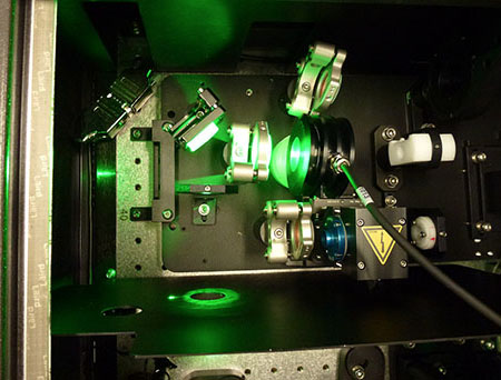
Photon Machines Analyte198 Femto-second laser ablation system.
JOHN DE LAETER CENTRE
The John de Laeter Centre houses a suite of mass spectrometry instruments and is a collaborative research venture involving Curtin University, the University of Western Australia, CSIRO and the Geological Survey of WA. It hosts over $25M in infrastructure in key facilities supporting research in: geosciences (geochronology, thermochronology and isotope studies); environmental science and global change; isotope metrology; forensic sciences; economic geology (minerals and petroleum); marine sciences; nuclear sciences. The components are organised into nine major facilities.
The Advanced Ultra-Clean Environment (ACE) Facility: This consists of a ~400m2 class 1000 containment space that houses four class 10 ultra-clean laboratories, a class 10 reagent preparation laboratory and a −18 °C class 10 cold clean laboratory, located at Curtin University. The extremely low ultimate particle counts are achieved with successive ‘spaces within spaces’ and HEPA (99.999% high efficiency particle arresting) filtration at each stage.
Inductively-Coupled Plasma Mass Spectrometry (ICPMS) Facility: This facility is located at UWA and consists of:
• TJA (VG/Fisons) PlasmaQuad 3 Quadrupole ICP-MS. The system has a high sensitivity interface to facilitate ultra-low detection limits.
• TJA (VG/Fisons) Laserlab high resolution 266 nm (Frequency quadrupled Nd-YAG) laser. The laser system is adapted with a high-resolution interface to facilitate the ablation of craters down to 10 μm in diameter.
• GBC Optimas 8000 Time of Flight ICP-MS
• Leco Renaissance Time of Flight ICP-MS
• A wide range of chromatographic and thermal dissociation interfacing is also available.
Argon Isotope Facility: This is located at Curtin and is equipped with a MAP 215-50 mass spectrometer with a low-blank automated extraction system coupled with a NewWave Nd-YAG dual IR (1064 nm) and UV (216 nm) laser, an electron multiplier detector and Niers source. Laser analysis allows for high spatial resolution up to 10 μm beam size for UV laser and 300 μm for IR laser. Larger sample sizes (>8-10 mg) are accommodated by an automated Pond-Engineering low-blank furnace. The extraction line has a Nitrogen cryocooler trap and three GP10 getters that allow gas purification. An Argus VI Mass Spectrometer and a Photon Machines Laser have been ordered for the JdL facility.
A joint ANU-John de Laeter Centre for Mass Spectrometry (JdL) Argon Facility has been established following a successful ARC bid. A total of ~$988,200 will be expended with the ARC contribution being $420,000. A management committee comprising two Facility Directors (Dr Marnie Forster, RSES, and
Dr Fred Jourdan, JdL), Director JdL (Professor Brent McInnes),
Mr Michael Avent (School Manager, RSES) and Professor Gordon Lister (RSES named Project Manager and Chief Investigator on the ARC grant) has been set up and approved by all Collaborating and Partner Organisations.
The new instrument allows the JDLC Argon isotope facility to carry out specialised work on rare extra-terrestrial sample materials, such as micrometer-size grains recovered from the Itokawa asteroid (see below) by the Japanese spacecraft Hayabusa. Dr Jourdan was awarded the grains because of the international standing of his laboratory in the study of meteorites and asteroid impacts, as well as the new Argus instrument established in the John de Later Centre for Isotope Research.
A new Thermo Argus VI multi-collector noble gas mass spectrometer was installed in November 2012 with funding from a 2012 ARC LIEF grant. This new instrument is a low volume (~700 cc) instrument providing excellent sensitivity and is particularly suited to the isotopic analysis of small samples of the noble gases, and in particular, Argon. The muti-collector design gives it the ability to measure all five Ar isotopes simultaneously leading to reduced analysis time and greater productivity.
Organic Geochemistry Facility: This facility is located within Applied Chemistry at Curtin and the instruments used for biomarker, petroleum and water studies include:
• GCMS (Gas Chromatograph Mass Spectrometer)
• GC-HRMS-MS (Gas Chromatograph-High Resolution Mass Spectrometer)
• Py-GC-MS (Pyrolysis-Gas Chromatograph-Mass Spectrometer)
• LCTOFMS (Liquid Chromatograph-Time of Flight-Mass Spectrometer)
• LC-MS-MS (Liquid Chromatography-Mass Spectrometry-Mass Spectrometer)
• HPSEC-DAD (High Performance Size Exclusion Chromatograph-Diode-Array Detection)
• GCIRMS (Gas Chromatograph-Isotope Ratio-Mass Spectrometer)
• TD-GC-IRMS (Thermal Desorption-Gas Chromatograph-Isotope Ratio-Mass Spectrometer)
• EA-IRMS (Elemental Analysis-Isotope Ratio-Mass Spectrometer)
Sensitive High Resolution Ion Micro Probe (SHRIMP):
The facility at Curtin has two automated SHRIMP II ion microprobes capable of 24-hour operation, together with a preparation laboratory. The equipment allows in-situ isotopic analysis of chemically complex materials with a spatial resolution of 5-20 microns. The main application of the SHRIMP instruments at Curtin is for U-Th-Pb geochronology of mineral samples. Zircon and other U-bearing minerals, including monazite, xenotime, titanite, allanite, rutile, apatite, badeleyite, cassiterite, perovskite and uraninite are the main minerals studied, where multiple growth zones commonly require high spatial resolution analyses.
Stable Isotope Ratio Mass Spectrometry (SIRMS) Facility: The West Australian Biogeochemistry Centre (WABC) at UWA is associated with the WA John de Laeter Centre of Mass Spectrometry and provides a range of analytical and interpretive services to researchers both within UWA and in the broader scientific community. The WABC currently operates three isotopic ratio mass spectrometers (IRMS) plus a considerable range of further analytical instrumentation (GC, HPLC, CE autoanalyser) routinely used in biogeochemical studies. A fourth IRMS (especially for small-sample δ13C and δ18O and carbonate analysis) is now being commissioned. Our IRMS are coupled with a variety of sample preparation modules to facilitate analysis of a broad range of sample matrices. Consequently, a wide range of applications of stable isotopes is supported by this facility.
Thermal Ionisation Mass Spectrometry (TIMS) Facility: The TIMS facility at Curtin incorporates a Thermo Finnegan Triton™ and a VG 354 multicollector mass spectrometer. The Triton is equipped with a 21-sample turret and 9 faraday cups, enabling a precision of 0.001% on isotopic ratios. As well as geological applications within the broad field of isotope geochronology (Re/Os, U/ Pb, Pb/Pb, Sm/Nd, Rb/Sr) the TIMS instruments can be applied to a variety of subject areas involving isotope fingerprinting, such as mantle geodynamics, forensics and the environmental impact of human activities. The TIMS instruments are also widely used in chemical metrology for the calibration of isotopic standards, and the calculation of isotopic abundances and atomic weights.
AuScope GeoHistory and (U-Th)/He Facility: The laboratory at Curtin hosts the prototype of the Alphachron™ automated helium microanalysis instrument now marketed by Australian Scientific Instruments in Canberra. (U-Th)/He thermochronology involves the measurement of 4He generated by the radioactive decay of U and Th in minerals. Helium is an inert gas that is quantitatively retained by minerals at low temperature, but is gradually lost from the mineral lattice by diffusion at elevated temperatures. Some minerals are more retentive to helium than others (e.g. zircon = 200 °C vs apatite = 75 °C), a unique characteristic that, when integrated with other techniques such as U-Th-Pb and Ar-Ar dating, can be used to produce complete time-temperature histories through a temperature interval from 900 °C to 20 °C. The JDLC (U-Th)/He Facility provides thermal-history analysis of metallogenic and petroleum systems by integrating several age-dating capabilities along with 4D thermal modelling. The Facility is also involved in fundamental collaborative research in the fields of orogenic tectonics, volcanology and quantitative geomorphology. The facility has grown in 2012-13 to integrate a new Alphachron™ machine coupled to a RESOlution Excimer laser + Agilent 7700 mass spectrometer. This “RESOchron” instrument enables the development of in-situ U-Pb and (U-Th)/He dating on single crystals of U-bearing minerals and immensely increases our application potential. In addition, laser ablation trace element analysis and U-Pb geochronology is now routinely undertaken in this facility, supporting industry, government and University projects.
K-Ar Geochronology Facility: The K-Ar facility utilises the following instrumentation and techniques:
• VG3600 noble gas mass spectrometer
• Heine double vacuum resistance furnace
• Clay mineral separation laboratory utilising cryogenic disaggregation of rock samples
• XRD, SEM, TEM, particle size analysis for clay characterisation
• Vacuum encapsulation station for Ar-Ar dating of ultrafine samples
• Clays – illite: Dating the timing of diagenetic and deformation events
• Fault gouge dating (illite) – earthquake and hazard assessment
selFRAG Facility: A new SelFRAG facility has been completed within the JDLC at Curtin University. The facility provides electric pulse dissagregation methodologies for mineral separation that allow pristine mineral grains to be separated from rock samples without the damage associated with standard crushing techniques.
Electron Microscopy Facility (EMF): The EMF is a new node of the John de Laeter Centre located at Curtin University and is a state of the art facility for microanalysis using electron microscopes. The facility has three scanning electron microscopes, all with EBSD capability, and a transmission electron microscope. The suite of instruments facilitates high quality research across a range of disciplines including Earth sciences, materials science, engineering, chemistry and biology.
The following is a summary of the capabilities of our major instruments;
Zeiss NEON 40 EsB - The Neon is a dual beam focused ion beam scanning electron microscope (FIB-SEM) equipped with a field emission gun and a liquid metal Ga+ ion source. This instrument combines high resolution imaging with precision ion beam ablation of focused regions which allows for site specific analysis of the surface and subsurface of samples in 2D or 3D.
Key Capabilities
• High resolution imaging using SE, BSE and inlens detectors (resolution is 1.1 nm at 20 kV to 2.5 nm at 1 kV)
• Energy Dispersive X-ray Spectroscopy (EDS) point analysis and mapping
• Electron Backscatter Diffraction (EBSD) mapping, including 3D EBSD
• Focused Ion Beam (FIB) milling
• Transmission Electron Microscope (TEM) lamella, Transmission Kikuchi Diffraction (TKD) foil and atom probe tip preparation
• High resolution 3D tomography
Tescan MIRA3 XMU with Oxford AZTEC EBSD-EDS system
The MIRA is a variable pressure field emission scanning electron microscope (VP-FESEM) equipped with a comprehensive range of detectors suitable for researchers in the fields of Earth science, forensics, life science and materials science.
Key Capabilities
• High resolution SE and BSE imaging of 1-3 nm (depending upon conditions used)
• Fast Electron Backscatter Diffraction (EBSD) mapping
• Energy Dispersive X-ray Spectroscopy (EDS) point analysis and mapping
• High quality cathodoluminescence (CL) imaging
• Electron Beam Induced Current (EBIC) imaging
• Low vacuum secondary electron imaging up to 500 Pa
• Scanning Transmission Electron Microscope (STEM) imaging
• Beam Deceleration Mode (BDM) for low voltage imaging of beam sensitive samples
• Simultaneous EBSD and EDS mapping
• Large area autonomous data collection
• Stereoscopic imaging
Zeiss EVO
The Zeiss EVO 40XVP is a variable pressure scanning electron microscope (VP-SEM) equipped with a tungsten filament. The microscope is suitable for general-purpose microstructural analysis at high vacuum, or for the analysis of non-conductive/hydrated samples at lower vacuum.
Key Capabilities
• Secondary Electron (SE) imaging
• Backscattered Electron (BSE) imaging
• Cathodoluminescence (CL) imaging
• Low vacuum secondary electron imaging
• Energy Dispersive X-ray Spectroscopy (EDS) point analysis and mapping
• Electron Backscatter Diffraction (EBSD) mapping
Jeol JEM 2011
The JEM-2011 is a transmission electron microscope (TEM) with a LaB6 filament. The TEM is equipped with an energy dispersive x-ray spectroscopy detector and a scanning transmission electron microscope attachment. This combination allows for comprehensive elemental and microstructural analysis at very high magnifications.
Key Capabilities
• High resolution bright field and dark field imaging
• Scanning Transmission Electron Microscope (STEM) imaging
• Selected Area Electron Diffraction (SAED)
• Energy Dispersive X-ray Spectroscopy (EDS) analysis
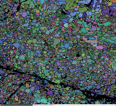
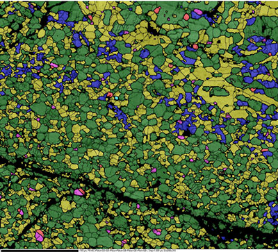
Phase map and an all-Euler orientation map of a basaltic achondrite meteorite. Thick black lines are phase boundaries, thin black lines are grain boundaries (same phase). The colours in the phase map are blue (quartz); yellow (anorthite); green (augite); brown (enstatite); aqua (olivine); fuchsia (ilmenite); red (chromite); orange (zircon). Black areas indicate no indexing and represent sample damage most likely associated with formation of the meteorite. Data was collected on the Tescan Mira XMU. Number of points (diffraction patterns) = 113,208. Collection time ~50mins (EBSD and EDS data collected).
|
|
WESTERN AUSTRALIA PALEOMAGNETIC AND ROCK-MAGNETIC FACILITY
The Western Australia Paleomagnetic and Rock-magnetic Facility was established at the University of Western Australia by CCFS CI Z.X. Li in 1990, funded by a UWA start-up grant to the late Professor Chris Powell. It was subsequently upgraded through an ARC Large Instrument Grant in 1993 to purchase a then state-of-the-art 2G Enterprises AC-SQUID cryogenic magnetometer and ancillary demagnetisation and rock magnetic instruments. It was upgraded again in 2006 into a regional facility, jointly operated by Curtin University, UWA and the Geological Survey of WA through an ARC LIEF grant with a 4k DC SQUID system plus a Variable Field Translation Balance (VFTB). A MFK-1FB kappabridge was installed in 2011.
The facility is one of the three similar laboratories in Australia, with major instruments including:
• 2G cryogenic magnetometer upgraded (LE0668377) to a 4K DC SQUID system
• MMTD80 (one) and MMTD18 (two) thermodemagnetisers
• Variable Field Translation Balance (VFTB)
• MFK-1FB kappabridge
• Bartington susceptibility meter MS2 with MS2W furnace
A wide range of research topics have been investigated using the facility, including reconstructing the configuration and drifting history of continents all over the world from the Precambrian to the present, analysing regional and local structures and deformation histories, dating sedimentary rocks and thermal/chemical (e.g. mineralisation) events, orienting rock cores from drill-holes, tracing ancient latitude changes, paleoclimates, and recent environmental pollution.
Program 1: Regional and Global Tectonic Studies
Paleomagnetism and rock magnetism are employed to study tectonic problems ranging from global to microscopic scales. The WA research group plays a leading role in a worldwide effort to establish the configuration and evolution of supercontinents Pangaea, Gondwanaland, Rodinia, and pre-Rodinia supercontinents.
Program 2: Ore genesis studies and geophysical exploration
We carried out a major research program on the timing and genesis of the giant iron ore deposits in the Pilbara region, and obtained a systematic set of petrophysical parameters for rock units in the region that enables more reliable interpretations of geophysical survey results (gravity and magnetic).
Program 3: Magnetic signatures in sediments as markers of environmental change
Sediments in suitable environments can incorporate a large number of environmental proxies. A major strength of environmental-magnetism analyses, such as magnetic susceptibility and saturated isothermal magnetism, is that they provide a rapid and non-destructive method of obtaining information on changes in paleoclimate and environment of sedimentation. In addition, rock magnetism can be used for monitoring and tracing industrial pollution.
Program 4: Magnetostratigraphy
We are conducting major research programs in the Canning Basin and in East Timor, both linked to petroleum resources.

 ARC Centre of Excellence for Core to Crust Fluid Systems