Infrastructure and technology development
CCFS links three internationally recognised concentrations of analytical geochemistry infrastructure: GEMOC’s Geochemical Analysis Unit (Macquarie University) and the associated Computing Cluster, the Centre for Microscopy, Characterisation and Analysis (UWA/Curtin) and the John de Laeter Centre of Mass Spectrometry. All are nodes for the NCRIS AuScope and Characterisation Capabilities, and have complementary instrumentation and laboratories. In addition, Curtin and UWA share a leading facility for paleomagnetic studies, and facilities for experimental mineralogy and petrology are being built up at Macquarie and Curtin.
Facilities:
CCFS/GEMOC INFRASTRUCTURE, LABORATORIES AND INSTRUMENTATION
CMCA TECHNOLOGY DEVELOPMENT AND INSTRUMENTATION
WESTERN AUSTRALIA PALAEOMAGNETIC AND ROCK-MAGNETIC FACILITY
CCFS/GEMOC INFRASTRUCTURE, LABORATORIES AND INSTRUMENTATION
CCFS links three internationally recognised concentrations of analytical geochemistry infrastructure: GEMOC’s Geochemical Analysis Unit (Macquarie University, reorganised in 2016 as MQGA) and the associated Computing Cluster, the Centre for Microscopy, Characterisation and Analysis (UWA/Curtin) and the John de Laeter Centre of Mass Spectrometry. All are nodes for the NCRIS AuScope and Characterisation Capabilities, and have complementary instrumentation and laboratories. In addition, Curtin and UWA share a leading facility for paleomagnetic studies, and facilities for experimental mineralogy and petrology are being built up at Macquarie and Curtin.
CCFS/GEMOC INFRASTRUCTURE, LABORATORIES AND INSTRUMENTATION
The analytical instrumentation and support facilities of MQ Geoanalytical represent a state-of-the-art geochemical facility.
The GAU contains:
- a Cameca SX-100 electron microprobe
- a Zeiss EVO MA15 Scanning electron microscope (with Oxford Instruments Aztec Synergy EDS/EBSD and Horiba HCLUE spectral cathodoluminescence detector)
- four Agilent quadrupole ICPMS (industry collaboration; two 7500cs; two 7700cx)
- two Nu Plasma multi-collector ICPMS (one decommissioned in June 2015)
- a Nu Plasma II multi-collector ICPMS (installed in June 2015)
- a Nu Attom high resolution single-collector sector field ICPMS
- a Thermo Finnigan Triton TIMS
- three New Wave laser microprobes (one 266 nm, two 213 nm, each fitted with large-format sample cells) for the MC-ICPMS and ICPMS laboratories (industry collaboration)
- two Photon Machines Excite Excimer laser ablation systems
- a Photon Analyte G2 Excimer laser ablation system
- a Photon Machines Analyte198 Femtosecond laser ablation system
- a PANalytical Axios 1kW XRF with rocker-furnace sample preparation equipment
- a Vario El Cube CHNS elemental analyser
- an Ortec Alpha Particle counter
- a New Wave MicroMill micro-sampling apparatus
- a ThermoFisher iN10 FTIR microscope
- a Horiba LABRAM HR Evolution confocal laser Raman microscope
- a selFrag electrostatic rock disaggregation facility
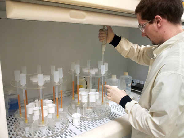
Romain Tilhac separating Sr, Nd and Hf by column chromatography for isotope analysis (Photo by Soumaya Abbassi).
Clean labs and sampling facilities provide infrastructure for ICPMS, XRF and isotopic analyses of small and/or low-level samples.
Experimental petrology laboratories currently include two piston-cylinder presses (pressures to 4 GPa), hydrothermal apparatus, Griggs apparatus and a multi-anvil apparatus for pressures to 27 GPa. Additional multi-anvil and piston-cylinder presses, plus a Laser-heated Diamond Anvil Cell apparatus are currently being acquired.
THE GEMOC FACILITY FOR INTEGRATED MICROANALYSIS (FIM) AND MICRO-GIS DEVELOPMENT
This facility was built up to fulfil the vision of providing spatially controlled high-resolution analysis and imaging of trace elements and isotopic abundances in situ, analogous to the capabilities of the electron microprobe for major elements in geological materials. This unique vision and approach enabled benchmark technology and in situ analytical methodology milestones in GEMOC starting with trace elements in mantle minerals from the mid-1990s, Hf isotopes in zircon from 2000, and Re-Os in mantle sulfides and alloys also from 2000. This distinctive in situ approach sparked research into new ways of understanding earth processes, and identified GEMOC, then CCFS, as the leading geochemical facility for such applications, and distinguished it from outstanding analytical laboratories that continued to undertake bulk analytical approaches. The new Decadal Plan for Earth Sciences prepared by the Australian Academy of Science National Committee of Earth Sciences has identified the continuation of in situ analysis as the preferred direction for geochemical analytical applications for industry and academia over the next 10 years.
This facility is based on in situ imaging and microanalysis of trace elements and isotopic ratios in minerals, rocks and fluids. The Facility for Integrated Microanalysis consists of four different types of analytical instrument, linked by a single sample positioning and referencing system to combine spot analysis with images of spatial variations in composition (‘micro-GIS’). The FIM has been in operation since mid-1999. Major instruments were replaced or upgraded in 2002-2004 through the $5.125 million DEST Infrastructure grant awarded to GEMOC, Macquarie University with the Universities of Newcastle, Sydney, Western Sydney and Wollongong as partners. Further enhancement of the facility took place following the award of an ARC LIEF grant in 2010 to integrate the two existing multi- collector inductively-coupled-plasma mass spectrometers (MC-ICPMS) with three new instruments: a femtosecond laser-ablation microprobe (LAM; installed in June 2012); a high-sensitivity magnetic-sector Nu Attom ICPMS (installed in January 2013); an Agilent 7700 quadrupole ICPMS (installed in 2010). In 2012 GEMOC was awarded ARC LIEF funding for a second generation MC-ICPMS and a Nu Plasma II was installed in June 2015.
The geochemical facilities operations at Macquarie University were reorganised by the Department of Earth and Planetary Sciences in 2016 without consulting CCFS, and input from CCFS during the subsequent GAU review process, to ensure operational security, was not taken into account. Staff changes in early 2016 resulted in a shortfall of three key positions (including the resignations of David Adams and Dr Will Powell). As a result, the functioning of some instruments (including the electron microprobe, the SEM and the LAM-ICPMS) was compromised throughout 2016. Research staff from CCFS were able to keep the SEM, LAM-ICPMS and multi-collector (MC) ICPMS in operation, producing CCFS-GEMOC’s unique suite of in situ results key to many CCFS projects. Despite the efforts of the CCFS staff, this series of problems and lack of experienced leadership impinged on the research projects of many staff and students.
Equipment for high-pressure experimentation
The expansion of the high-pressure experimental facilities continued, with plans for an extension to the laboratory in a new wing extending into the yard behind building E5A. This will house two large multi-anvil presses, and a third will be installed in the current laboratory. Laser-heated diamond anvil cells and an additional piston-cylinder apparatus are being acquired through LIEF funds from the Australian Research Council. An experimental program on electrical conductivity in mantle materials has begun with the currently available multi-anvil apparatus, and experiments studying the reaction between melts of sediment and mantle peridotite are being conducted.
PROGRESS IN 2016:
1. Facility for Integrated Microanalysis
a. Electron Microprobe: As noted above, the electron microprobe laboratory did not produce results throughout 2016, due to the staffing situation and the lack of appropriate technical expertise. As a stopgap measure, CCFS PhD students and research staff were directed to other microprobe laboratories nationwide and internationally. We are grateful to our colleagues at these institutions (among others) University of Melbourne, ANU-RSES, University of Western Sydney, University of New South Wales, Mainz University and IGGCAS, Beijing (a CCFS international Partner Organisation), for their expert and enthusiastic assistance for instrument access. Sarah Gain is especially thanked for her efforts in keeping the SEM operative and available to students until the Department, late in 2016, provided a part-time student assistant to help with the SEM. The EMP is not yet operational due to lack of care during downtime which resulted in failing pumps and detectors. Dr Timothy Murphy has been appointed to oversee the electron microprobe functions, and arrives in early 2017.
CCFS has provided significant funding support and scientific expertise to purchase a Scanning X-ray spectrometer to enable fast scanning and mapping of thin sections and blocks thus providing a wider and more complete spatial framework for in situ analysis. The acquisition and running of this instrument is a joint venture with Prof Damien Gore (Department of Geography). The versatility of this instrument has attracted a significant interest from most faculties across Macquarie University, including Arts.
b. Laser-ablation ICPMS microprobe (LAM): .
In 2016 the combination of the Photon Machines G2 laser system and Agilent 7700 ICPMS was used for in situ trace element analyses and U-Pb geochronology. The facility was used by 12 Macquarie PhD thesis projects, 6 international visitors, 6 Masters Research students, 15 users from other Australian institutions and several in-house funded research projects and industry collaborations. Projects included the analysis of minerals from mantle-derived peridotites, pyroxenites and chromitites, new, unusual types of ultra-reduced phases from volcanic sources and ultra-high pressures terranes, high-grade metamorphic rocks and biominerals.
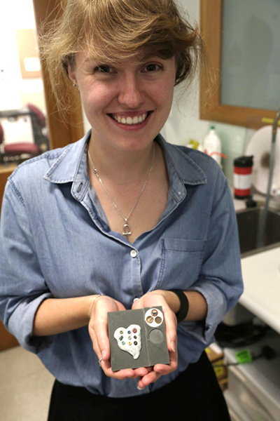
Archaeologist Michelle Whitford with Egyptian beads for LAM analysis.
More unusual materials were also analysed including Egyptian pottery and stone beads and remains of swords from the Dubai area, in collaboration with Ivan Stepanov and Prof Lloyd Weeks (UNE). Further development regarding the fingerprinting of archaeological materials is in progress and discussions are underway with the Department of Ancient History.
Following the resignation of Dr Will Powell in early 2016, U-Pb analysis of zircon was enabled through 2016 by the expertise of CCFS Research Associates Rosanna Murphy and Yoann Gréau with generous input from other CCFS researchers. Sarah Gain and Rosanna Murphy also kept the laboratory operating for the analysis of trace elements in a variety of materials. TerraneChron®(http://www.gemoc.mq.edu.au/TerraneChron.html) continued. Method development and TerraneChron®(http://www.gemoc.mq.edu.au/TerraneChron.html) work were restricted in 2016 due to the reorganisation, staffing shortfall, and resulting difficulties in maintaining instrument operation.
A Technical Officer to provide technical expertise has been selected and is likely to arrive in May 2017.
c. MC-ICPMS: A Nu Plasma II MC-ICPMS was installed in June 2015 and followed the decommissioning of Nu Plasma 005, after 16 years of service. Although the Nu Plasma II represents a significant advance in its electronics and engineering, much of the fundamental design is adapted from Nu Plasma I. This enabled a relatively seamless transition of existing methods developed over the past 15 years on the Nu Plasma I. The combination of the expanded collector array (16 Faraday cups and 5 ion counters) and enhanced sensitivity compared to the first generation Nu Plasma instruments has enabled the refinement of several in situ techniques pioneered at GEMOC, Macquarie.
The in situ measurement of U-Pb isotopes in zircon using the combination of the femtosecond laser system and Nu Plasma II was a world first, with preliminary results reported at the Goldschmidt Conference in Prague, August 2015 (N.J. Pearson, W.J. Powell, Y. Gréau, R.C. Murphy, J.L. Payne, E. Belousova, W.L. Griffin and S. Y. O’Reilly 2015. U-Pb geochronology of zircon by femtosecond laser ablation, Goldschmidt Abstracts, 2015, 2437). The development of standard operating procedures for in situ U-Pb, Re-Os and Rb-Sr isotope measurements is on-going. The development of Mg isotope methodologies for chromite and chromite-rich ultramafic rocks as part of the TARDIS Program (Nicole McGowan PhD) in the ARC Centre of Excellence for Core to Crust Fluid Systems (CCFS) was completed; high-precision results were obtained in wet-plasma mode on the Nu Plasma II and corroborated by replicate measurements at IGGCAS, Beijing.
At the time of the installation of the new Nu Plasma II, Nu Plasma HR 034 underwent an upgrade with an enhanced interface. The upgrade increased sensitivity between 1.5 and 2 times, and this contributed to an overall improvement in signal stability, as well as in the precision of single measurements and long- term reproducibility. In 2015 a third Photon Machines excimer laser microprobe was installed and co-located with Nu Plasma HR 034. After successful installation and commissioning had been achieved and first experiments with the Femtosecond laser microprobe were completed (and presented at the 2015 Goldschmidt conference), key staff to develop the integrated system and applications were no longer available in 2016.
A new Future Fellow, Dr Olivier Alard (pictured), has relevant expertise and a technical position replacement process is in progress. In addition, the ARC Centre of Excellence for Core to Crust Fluid Systems has funded a technology development program employing a Research Associate (Dr Yoann Gréau) with a high level of instrument expertise who, with Dr Alard, has been recently making good progress with the envisaged developments. A new split-stream approach is now being investigated, involving additional gas lines and mass-flow controllers to control the amount of aerosol transported into each instrument. Ultimately, this approach will assist in achieving adequate sensitivity on both sides of the system, and therefore will optimise both signal outputs for a given ablated volume. The planned first application of this new methodology will be combined U-Pb and Lu-Hf characterisation of zircons and simultaneous measurements of Pb-Pb and Re-Os in sulfides.
The LAM MC-ICPMS is the vehicle to deliver in situ high-precision ratio measurements including the analysis of Lu-Hf isotopes in zircon as a major part of TerraneChron®. CCFS/GEMOC remains one of the few facilities with the capability to perform in situ Re-Os dating of single grains of Fe-Ni sulfides and alloys in mantle- derived rocks. Re-Os studies were undertaken on xenoliths from eastern China, Siberia, Italy and Algeria (Hoggar), and sulfide and platinum group minerals in chromitites from Tibet, Australia, Spain and Turkey. This activity was also made possible by the expertise of CCFS Research Associates Rosanna Murphy and Yoann Gréau with input from other CCFS researchers, and CCFS Future Fellow Olivier Alard, following the resignations in early 2016. CCFS is translating the full methodology to the Taiwan Academia Sinica laboratories under the auspices of Dr Kuo-Lung Wang, a former GEMOC Research Fellow and CCFS Associate Researcher, to ensure preservation of the associated intellectual property and approach.
d. Laboratory development: The clean-room facility established in 2004 continued to be used primarily for isotope separations for analysis on the Triton TIMS and Nu Plasma MC-ICPMS. Routine procedures continued for Rb-Sr, Nd-Sm, Lu-Hf and Pb isotopes, as well as U-series methods (U, Th and Ra).
e. Software: GLITTER (GEMOC Laser ICPMS Total Trace Element Reduction) software is our on-line interactive program for quantitative trace element and isotopic analysis and features dynamically linked graphics and analysis tables. This package provides real-time interactive data reduction for LAM-ICPMS analysis, allowing inspection and evaluation of each result before the next analysis spot is chosen. GLITTER’s capabilities include the on-line reduction of U-Pb data. Sales of GLITTER are handled by AccessMQ and GEMOC provides customer service and technical backup. During 2016 a further 16 full licences of GLITTER were sold, bringing the total number in use to more than 287 worldwide, predominantly in Earth sciences applications but with growing usage in forensics and materials science.
Dr Will Powell continued in his role in GLITTER technical support and software development through 2016 on a consultancy basis, following his resignation and relocation to Rio Tinto (Melbourne) in early 2016. The current GLITTER release is version 4.4.4 and is currently available without charge to existing customers.2. X-Ray Fluorescence Analysis
In November 2012 a PANalytical Axios 1 kW X-ray Fluorescence Spectrometer was installed and is used routinely to measure whole-rock major element compositions on fused glass discs and trace-element concentrations on pressed-powder pellets. In 2013 the sample preparation equipment was upgraded and included a new furnace to make high-quality cast glass beads. The major element calibration was modified in 2015 to extend the spectrum of rock types that could be analysed to include Fe-rich samples such as iron ores and laterites.
2.1. A high performance CHNS elemental analyser from Elementar (Vario El Cube) fitted with an extra IR-detector for low-level sulfur analysis has been purchased and recently installed. This facility enables us to better estimate whole-rock sulfur contents and thus sulfide abundance in order to select appropriate samples for in situ Re-Os isotopic analysis.
3. Whole-rock solution analysis
An Agilent 7500cs ICPMS produces trace-element analyses of dissolved rock samples for the projects of CCFS/GEMOC researchers and students and external users, supplementing the data from the XRF.
The ICPMS dedicated to solution analysis is also used to support the development of ‘non-traditional’ stable isotopes with the refinement of separation techniques and analytical protocols (see 1. d).4. Diamond preparation and analysis
The GEMOC laser-cutting system (donated by Argyle diamonds in 2008) was used through 2015 to cut thin plates of single diamond crystals as part of the on-going research into diamond genesis. However, in 2016 the instrument has repeatedly broken down and is being decommissioned.
5. selFrag - a new approach to sample preparation
GEMOC’s selFrag instrument was installed in May 2010 and was the first unit in Australia. This instrument uses high-powered electrical pulses to disaggregate rocks and other materials along the grain boundaries. It removes the need to crush rocks for mineral separation, and provides a higher proportion of unbroken grains of trace minerals such as zircon. Since its installation selFrag has been used for a range of applications including zircon separation, the analysis of grain size and shape in complex rocks, and the liberation of trace minerals from a range of mantle-derived and crustal rocks.
Sarah Gain with personnel from Horiba and Quark Photonics during the installation of the CL monochromator on the SEM.
6. Spectroscopy
The spectroscopy infrastructure includes an FTIR microscope (ThermoFisher iN10 FTIR microscope; 2008). The FTIR is used to measure H abundance in a range of nominally anhydrous minerals (e.g. olivine, pyroxene, garnet) and H and N contents in diamond. In developing the spectroscopy capability an emphasis has been placed on hyperspectral mapping to produce integrated datasets and multi-layered information in a spatial context. A Horiba H-CLUE CL monochromator was installed on the Zeiss EVO SEM in January 2016. The monochromator system provides spatially resolved quantitative cathodoluminescence spectra, which allow identification of emitters (e.g. REE in zircons), crystal lattice vacancies (e.g. in diamond) and crystallographic information on how specific elements are incorporated in the mineral crystal lattices (e.g. Mn in aragonite). The new instrumentation is in the process of acquiring a growing group of users and is currently part of projects in biomineralisation (HDR student Laura Otter/Prof Dorrit Jacob), diamond growth (Professor Dorrit Jacob) and zircon characterisation (Honorary Associate Dr Christoph Lenz/ Dr Elena Belousova).
7. Raman spectrometry
A confocal laser Raman microscope (co-funded by MQSIS 2014 and Future Fellowship funding to Professor Dorrit Jacob) delivers information for non-destructive phase-identification and -characterisation at one micrometre spatial resolution.
The Raman spectrometer continues to serve the CCFS, the Department and the Faculty. This year the system’s capabilities were extended with the purchase of two new laser wavelengths (MQSIS 2017), upgrading the Raman spectrometer to one of the most versatile and capable in the Sydney area. The instrument enjoys attention from a growing user group across the Faculty of Science and Engineering at Macquarie University with users from Chemistry, Physics, Biology and Environmental Sciences as well as continued popularity with Dr Christoph Lenz from ANSTO. Professor Lutz Nasdala (University of Vienna), one of the world- leading experts in Raman Spectrometry provided a short course on Raman spectrometry for CCFS in December 2016.
In 2017 we plan to extend the applications of Raman Spectrometry towards forensics applications, namely ink characterisation on Egyptian papyrus (with Prof Damian Gore and Assoc Prof Malcolm Choat).
8. Computer cluster
The cluster Enki has continued to be a powerhouse for the geodynamics group, having supported multiple research projects, > 5 PhD projects, postdocs, and numerous Masters-level projects. Recent developments have included the incorporation of melt transport and crustal creation into the mantle convection code Aspect (based on the deal.II finite element libraries), led by Siqi Zhang. Other codes utilising Enki for simulation include O’Neill and Zhang’s smoothed-particle hydrodynamic codes to simulate early solar-system processes (currently in review). In addition, the lithosphere/seismic cluster “Toto” (managed by J.C. Afonso) continues operation, while a GPU Tower (supplied by Xenon systems) acts as a development machine for GPU-capable code, including the SPH code. A Xeon-Phi server (supplied by Dell) has recently been installed, enabling the modelling group to start development and migration of their codes onto this next generation hardware.
CMCA TECHNOLOGY DEVELOPMENT AND INSTRUMENTATION
The University of Western Australia’s Centre for Microscopy, Characterisation and Analysis (CMCA) is a $50 M core facility providing analytical solutions across a diverse array of scientific research. The world-class facilities and associated technical and academic expertise are the focus of micro-analytical and characterisation activities within Western Australia, while strong links and collaborations have earned the CMCA an excellent national and international reputation. The CMCA incorporates the Western Australian Centre for Microscopy, and is a node of the NCRIS Characterisation capabilities, the National Imaging Facility (NIF) and the Australian Microscopy and Microanalysis Research Facility (AMMRF). It is also associated with the NCRIS funded Australian National Fabrication Facility (ANFF), and AuScope, which have made a substantial contribution to facilities run by CMCA.
CMCA capabilities:
- Secondary Ion Mass Spectrometry (CAMECA IMS 1280 and CAMECA NanoSIMS 50 and NanoSIMS 50L)
- Electron probe microanalysis (JEOL JXA 8530F)
- Focused ion beam (FEI Helios)
- Transmission electron microscopy (FEI Titan, JEOL 2100)
- Scanning electron microscopy (FEI Verios XHR, Zeiss 1555, Tescan Vega3)
- X-ray powder diffraction (Panalytical Empyrean)
- X-ray micro-CT (Xradia)
- Confocal Raman imaging with AFM (WiTec Alpha 300RA+)
- NMR spectroscopy (2 Bruker Avance and 2 Varian spectrometers)
- X-ray crystallography (Oxford Diffraction)
- GC and HPLC mass spectrometry
- Bioimaging, flow cytometry, cell sorting, and laser micro-dissection
- Optical and confocal microscopy
- Biological sample cryo-preparation and ultramicrotomy
THE AMMRF FLAGSHIP ION PROBE FACILITY
The CAMECA IMS1280 and NanoSIMS 50 are flagship instruments of the AMMRF. The AMMRF Flagship Ion Probe Facility offers state-of-the-art secondary ion mass spectrometry (SIMS) capabilities to the Australian and international research communities, allowing in situ, high-precision isotopic and elemental analyses, and secondary ion imaging on a wide range of samples.
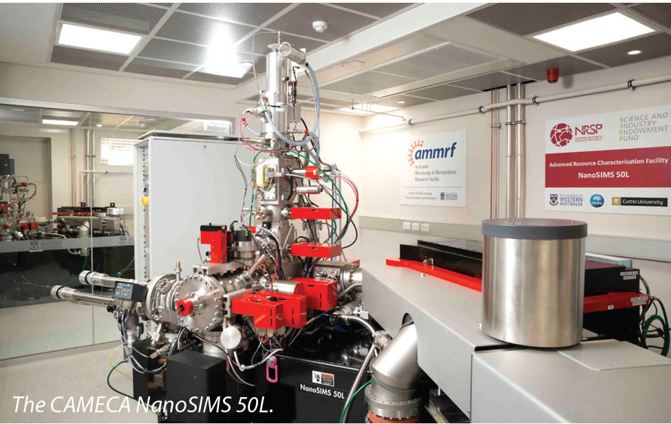
The IMS1280 large-geometry ion probe, installed in 2009, was co-funded by the University, the State Government of Western Australia, and the Federal Government’s Department of Innovation, Industry, Science and Research (DIISR) under the “Characterisation” (AMMRF) and “Structure and Evolution of the Australian Continent” (AuScope) capabilities of the National Collaborative Research Infrastructure Strategy (NCRIS). The NanoSIMS 50, installed in 2003, was funded through the Federal Government’s NCRIS-precursor, the Major National Research Facility scheme (NANO-MNRF). In 2015, the CMCA installed a new CAMECA NanoSIMS 50L ion probe as part of the NRSP’s Advanced Resources Characterisation Facility (ARCF). The ARCF provides multiscale characterisation capabilities for Geoscience research, from the scale of drill core down to atom scale. The Facility, funded through CSIRO’s Science and Industry Endowment Fund (SIEF), features the Geoscience Atom Probe installed in the John de Laeter Centre at Curtin University, and the MAIA mapping facility currently under development at CSIRO. UWA’s Ion Probe Facility can currently lay claim to being the best-equipped SIMS lab in the world, as no other facility has two NanoSIMS alongside an IMS1280.
UWA MSc student Bataar Bataar using the Cameca 120 ion probe for oxygen isotope analysis.
The Ion Probe Facility is a key characterisation component within the ARC Centre of Excellence for Core to Crust Fluid Systems. To ensure the highest levels of quality and throughput, CCFS has provided funding for a Research Associate position within the Ion Probe Facility, to facilitate direct scientific and technical interaction for all CCFS users and projects.
Progress IN 2016:
The Ion Probe Facility has continued to contribute to various projects in the context of CCFS. The CAMECA IMS1280 clocked up more than 4300 hours, far in excess of the ‘full-utilisation target’ of 1800 hours. The lab contributed to 42 individual projects, originating from CCFS partners, other Australian research institutes, and overseas. In addition, 7 new development projects were initiated to extend the lab’s capabilities, including REE measurements and ion imaging. 8 journal articles were published in 2016 featuring data acquired using the IMS1280 at UWA, of which 7 were directly related to CCFS projects.
With the new CAMECA NanoSIMS 50L coming online in late 2015, the NanoSIMS lab saw a big increase in the number of projects - 47, spread across 6500 hours. Projects originated from 11 Australian institutions, with 5 projects from overseas. Several new development projects that utilise the new RF plasma oxygen ion source were initiated, focusing predominantly on isotope mapping in minerals. The NanoSIMS lab contributed to 13 publications in 2016, of which 6 were Earth science-related. One high impact paper (Reith et al. Nature Geoscience) featured NanoSIMS imaging of Pt to shed light on the role of microbes in the transformation of PGE minerals. The CCFS pilot project “Making the invisible visible” yielded its first publication, illustrating the role of fluids in simplectite formation revealed by isotope labelling (CCFS Publication #866, Spruzeniece et al., J. Metamorphic Geol.).
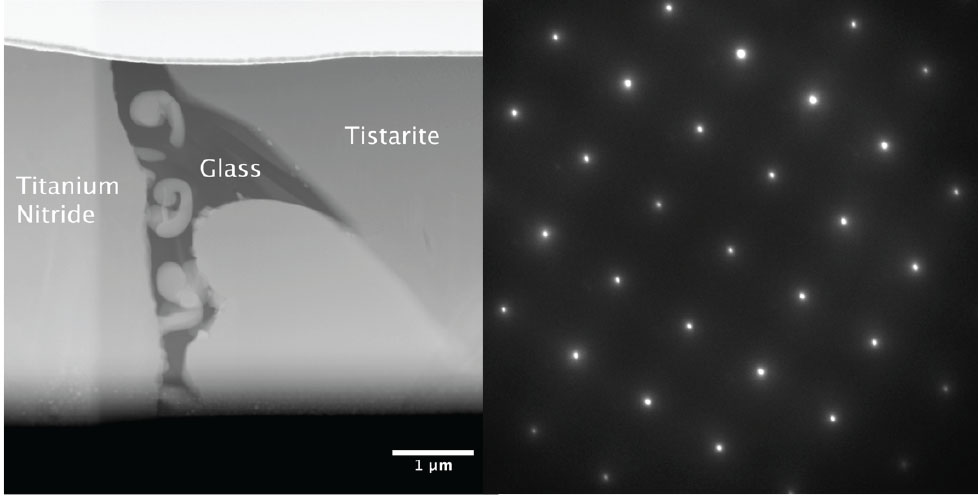
High-Angle Annular Dark Field STEM image (left) and diffraction pattern from the Tistarite phase (right).
CCFS research has also incorporated other analytical techniques at the CMCA. In 2016, the FEI Helios DualBeam and FEI Titan TEM were used to investigate the detailed mineralogy of ‘nebular’ mineral assemblages initially described in the 2015 CCFS Annual Report. In a collaboration between Bill Griffin’s Macquarie group and the CMCA’s Associate Prof Martin Saunders, the high resolution analytical capabilities of the Titan TEM have allowed the composition and crystal structure of these complex, fine-grained materials to be investigated, which has led to the identification of several exceedingly rare or previously unreported minerals. The first publication from the collaboration appeared in Geology and was featured as a ‘Nature Research highlight’ (CCFS Publication #830, Griffin et.al. 2016, Geology).
CMCA-CCFS 2016 publications were published in high profile journals such as Earth and Planetary Science Letters, Chemical Geology, Geological Society Special Publications, Journal of Metamorphic Geology and Terra Nova: CCFS Publications #797, 816, 830, 838, 847, 866, 921.
For further information on CMCA facilities please consult
http://www.cmca.uwa.edu.au/
JOHN DE LAETER CENTRE
The John de Laeter Centre (JdLC) is a collaborative research venture involving Curtin University, the University of Western Australia, CSIRO and theGeological Survey of Western Australia.
It hosts over $28M in infrastructure supporting research in: geosciences (geochronology, thermochronology and isotope studies); environmental science;isotope metrology; forensic science; economic geology (minerals and petroleum); marine science; and nuclear science.
The mission of the Centre is to “build world-class research infrastructure in Western Australia for the benefit of Earth, Environment and Materials Science research”. The JdLC isheadquartered in the Faculty of Science and Engineering at Curtin University, but has a governing board consisting of members of the joint venture partnersas well as representatives from the mining, petroleum and environment sectors.
The Centre experienced rapid growth in 2015 through the merger of mass spectrometry and microscopy facilities at Curtin, the commissioning of $5,200,000 innew analytical instrumentation and the appointment of 4 new research fellows. A new website has been developed to provide detailed information on the newfacilities, instrumentation and research staff (http://www.jdlc.edu.au).
The components of the JDLC are organised into fourteen major facilities including:
(GAP) Geoscience Atom Probe Facility:
GAP is a node of the Advanced Resources Characterisation Facility (ARCF) funded by a $12,400,000 Science and Industry Endowment Fund grant to Curtin, UWA and CSIRO. The GAP hosts a Cameca LEAP 4000X HR microscope capable of carrying out atom probe tomography (APT), a recent development in the geosciences, that provides high spatial resolution with time- of-flight mass spectrometry to provide 3-dimensional chemical information at the atomic scale. More commonly used to study semiconductors and metal alloys, the GAP is the first atom probe facility in the world to be dedicated to the study of geological materials (http://www.geoscienceatomprobe.org). The ARCF also commissioned a Tescan Lyra focused ion beam scanning electron microscope (FIB-SEM), with a Ga+ gun capable of micro- milling out a 100 nm wide needle of mineral sample prior to APT analysis. The Lyra system is a highly advanced platform for 2D and 3D microanalysis with time of flight mass spectrometry (TOF-SIMS) and electron back scattered diffraction (EBSD) detectors. By correlating the analytical outputs of both the LEAP and the Lyra instruments, the ARCF provides an unprecedented capability of characterising highly complex materials on a wide range of length scales.
(DMH) Digital Mineralogy Hub Facility:
The Facility hosts a Tescan Integrated Mineral Analyzer (TIMA GM) - a fully automated, high throughput, analytical Field Emission Gun Scanning Electron Microscope (FEGSEM) for automated analysis of sample composition. TIMA measures mineral abundance, liberation properties, mineral association and grain size automatically on multiple samples of grain mounts, thin sections or polished sections. Applications include ore characterisation, process optimisation, remediation and the search for precious metals and strategic elements. The facility is being used by a broad spectrum of researchers: geologists and archaeologists are using the facility in petrological characterisation, sample classification and lithofacies studies; while geochemists and geochronologists are using the mineral classification outputs as targeting maps for further ion, electron or laser microprobe analysis.(CEG) Curtin Experimental Geochemistry Facility:
CEG provides facility for experimental petrology, geochemistry and hydrogeochemistry at pressures and temperatures that range from those at the Earth’s surface to those at the base of the Earth’s crust. The Facility contains:
- 2 x 150 ton end loaded piston cylinder presses
- Coretest hydrothermal apparatus
- Assorted furnaces to 1400 degrees C
- Assorted titanium and Teflon-lined bombs
(GHF) GeoHistory Facility:
The GHF houses state-of-the-art laser ablation inductively coupled plasma mass spectrometry (LA-ICPMS) equipment, in addition to a low temperature thermochronology laboratory. The LA-ICPMS comprises a Resonetics S-155-LR 193nm excimer laser ablation system coupled to an Agilent 7700x quadrupole ICPMS. The Excimer laser is also coupled to a RESOchron helium analysis line for in situ (U-Th-Sm)/He, U-Pb and trace element analysis of single crystals. The facility also has a separate Alphachron helium line with a diode laser and furnace in order to facilitate conventional (U-Th)/He dating on single mineral crystals and larger samples. A Nu Plasma II multi-collector was integrated into the facility to facilitate split stream analysis.
(MMF) Microscopy and Microanalysis Facility:
The MMF houses a broad range of advanced microanalysis instrumentation providing high quality chemical, mineralogical and microstructural information, and high resolution images for research and technical publications. The facility staff have expertise in Materials and Earth Science research which is used to support both academic research and applied projects for the Western Australian minerals and energy sector. Techniques and instrumentation available include:
- High resolution imaging (TEM) - A new FEI Talos F200X S/TEM system will be commissioned in early 2017 to complement ongoing research at the nanoscale. The system combines high resolution S/TEM and TEM imaging with EDS and 3D chemical characterisation.
- The JEM is a transmission electron microscope (TEM) with a LaB6 filament. The TEM is equipped with an EDS detector and a scanning TEM attachment. This instrument is capable of elemental and microstructural analysis at extremely high magnifications.
- Spatially resolved elemental analysis (EDS) and Phase & orientation analysis (EBSD) - The MIRA3 is a variable pressure field emission scanning electron microscope (VP-FESEM) that features sensitive EDS and EBSD detectors and integrated software for high quality microstructural analysis of crystalline samples.
- Quantitative mineral analysis (Q-XRD) - The D8A is an X-ray Diffractometer (XRD) with a copper X-ray source and an automated 45 position sample changer. It features a LynxEye position sensitive detector that is 200 times faster than a conventional scintillator detector, allowing collection of superior data in a short time-frame.
- Ion beam sample manipulation including TEM & TKD lamella preparation (FIB) - The NEON is a dual beam focused ion beam scanning electron microscope (FIB-SEM) equipped with a field emission gun and a liquid metal Ga+ ion source. This instrument combines high resolution imaging with precision ion beam ablation of focused regions, allowing for site specific analysis of the surface and subsurface of samples in 2D or 3D.
- The MMF also houses a suite of equipment that includes light microscopy, vacuum mount impregnation, manual and
- automated polishers, mills and coaters that are used to prepare samples for electron microscopy and X-ray diffraction.
(SAXS) Small Angle X-Ray Scattering Facility:
Small angle X-ray scattering can be used to characterise the size, shape and distribution of objects between 1 and 100 nm. Instrumentation includes a Bruker NANOSTAR SAXS comprising a copper sealed tube X-ray source with a gas filled two dimensional photon counting detector. In 2016, LIEF funding was used to upgrade the instrumentation in the facility.
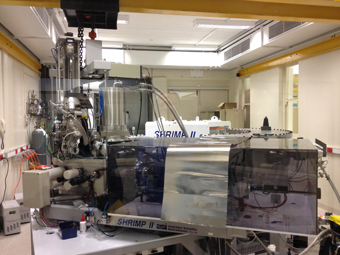
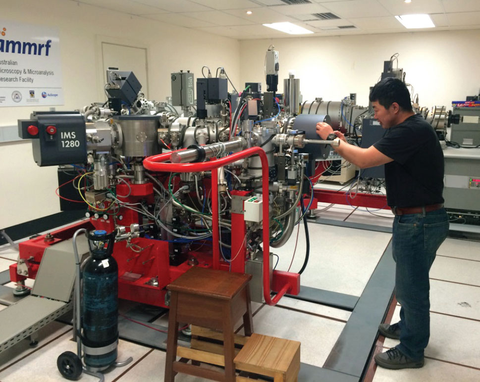
(SHRIMP) Sensitive High Resolution Ion Micro Probe Facility: The facility at Curtin has two automated SHRIMP II ion microprobes capable of 24-hour operation, together with a preparation laboratory that was remodelled in 2014. The equipment allows in situ isotopic analysis of chemically complex materials with a spatial resolution of 5-20 microns. The main application of the SHRIMP instruments at Curtin is for U-Th-Pb geochronology of zircon and other U-bearing minerals, including monazite, xenotime, titanite, allanite, rutile, apatite, baddeleyite, cassiterite, perovskite and uraninite where multiple growth zones commonly require analyses with high spatial resolution. SHRIMP II (pictured) is fitted with a Cs source, electron gun and 5 channel M/C. SHRIMP II A is currently being developed for stable isotope analysis of O in zircon and other silicates, and S in sulfides.
(SMS) SelFrag & Mineral Separation Facility:
A SelFrag facility, supported by an ARC LIEF grant, has been installed within the Department of Applied Geology at Curtin University. The facility provides electric pulse disaggregation for mineral separation, which allows mineral grains to be separated from rock samples without the damage associated with standard crushing techniques.
(TIMS) Thermal Ionisation Mass Spectrometry Facility:
The TIMS facility at Curtin incorporates a Thermo Finnegan Triton™ and a VG 354 multicollector mass spectrometer. The Triton is equipped with a 21-sample turret and 9 faraday cups, enabling a precision of 0.001% on isotopic ratios. As well as geological applications within the broad field of isotope geochemistry (Re/Os, U/ Pb, Pb/Pb, Sm/Nd, Rb/Sr) the TIMS instruments can be applied to a variety of isotope fingerprinting, such as forensics and the environmental impact of human activities. The TIMS instruments are also used for the calibration of isotopic standards and the calculation of isotopic abundances and atomic weights. The facility has recently installed a Thermo Scientific Triton™ mass spectrometer, facilitating a new range of geochemical, geological and environmental research applications.
(TRACE) TRACE Research Advanced Clean Environment Facility:
This consists of a ~400 m2 class 1000 containment space housing four class 10 ultra-clean laboratories, a class 10 reagent preparation laboratory and a −18 °C class 10 cold clean laboratory, located at Curtin University. The extremely low ultimate particle counts are achieved with successive ‘spaces within spaces’ and HEPA filtration at each stage.
(WAAIF) Western Australian Argon Isotope Facility:
This is located at Curtin and is equipped with A MAP215-50 mass spectrometer with a low-blank automated extraction system coupled with a New Wave Nd-YAG dual IR (1064 nm) and UV (216 nm) laser, an electromultiplier detector and Niers source. The ultra-violet laser is capable of high-resolution (up to 10 µm beam size) ablation of any mineral, allowing detailed analysis of individual mineral grains. The facility also houses an Argus VI Multi-Collector Noble Gas Mass Spectrometer.
The 40Ar/39Ar method is used to date a myriad of geological events such as volcanism, tectonic plate movements, mountain building rates, sediment formation, weathering and erosion, hydrothermal fluid movements, and alteration and diagenesis of minerals.
(WA-OIG) WA Organic and Isotope Geochemistry Facility:
WA-OIG is an internationally-recognised group contributing to world-class research in the fields of organic and stable isotope geochemistry, paleogenomics and geomicrobiology. Available techniques are listed here: http://jdlc.edu.au/wa-organic-and- isotope-geochemistry-facility-wa-oig/.
For further information on JDLC facilities please consult
http://www.jdlc.edu.au
WESTERN AUSTRALIA PALEOMAGNETIC AND ROCK-MAGNETIC FACILITY
The Western Australia Paleomagnetic and Rock-magnetic Facility, recently upgraded and relocated to Curtin University’s Bentley campus, is a national research infrastructure with the latest upgrade co-funded by the Australian Research Council and collaborating institutions including Curtin University, the University of Western Australia (UWA), the Australian National University, Macquarie University and University of Queensland. The facility was established at UWA in 1990 by CCFS CI Z.X. Li, and has been progressively upgraded over the years.
The latest upgrade includes the construction of a magnetically shielded room in mid-2015 by Dr Gary Scott’s team, which provides a laboratory space with ambient magnetic fields less than 0.5% of the local geomagnetic field. Within this shielded room we now have a new 2G 755 superconducting rock magnetometer with a vertical Model 855 automated sample handler (the RAPID system) and other accessories attached to it (automated AF demagnetiser, susceptibility meter, etc.). The RAPID system, the first and only one in Australia, was installed and commissioned in February 2017. Other systems now operating inside the shielded room include an AGICO JR-6A spinner magnetometer and ASC TD-48SC and MAGNETIC MEASUREMENTS thermal demagnetisers. The day-to-day operation of the facility is overseen by the recently appointed Technical Officer, Dr Josh Beardmore. A number of key pieces of apparatus are currently being relocated from UWA to the new Curtin precinct.
The new purchases represent a major enhancement to the productivity and capabilities of the facility. Apparatus which is now available in the facility include:
- a 2G 755 superconducting rock magnetometer with a vertical Model 855 automated sample handler (the RAPID system) and other accessories (including; AF coils, susceptibility meter, and ARM system)
- a second 2G 755 cryogenic magnetometer upgraded (LE0668377) to a 4K DC SQUID system (currently returned to 2G enterprises for a minor upgrade and for repair of the lightning-damaged cold head)
- An AGICO JR-6A spinner magnetometer
- 1x MMTD80, 2x MMTD18 and a TD-48-SC thermal demagnetiser
- a Petersen Instruments Variable Field Translation Balance (VFTB)
- an AGICO MFK-1FA kappabridge
- a Bartington MS2 susceptibility meter with MS2W furnace
- a MAGNETIC MEASUREMENTS MMPM5 pulse magnetiser
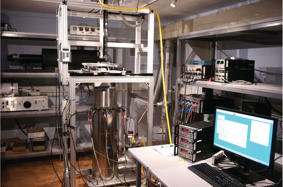
The 2G RAPID superconducting magnetometer system.
The facility supports a wide range of research topics, including reconstruction of global paleogeography (the configuration and drifting history of continents) through Earth’s history, studying the evolving geomagnetic field (e.g. paleointensity) through time, analyses of regional and local structures and tectonic histories, dating sedimentary rocks and thermal/chemical (e.g. mineralisation) events, studying past climate changes, and orienting rock cores from drill-holes.