Infrastructure and technology development
CCFS links three internationally recognised concentrations of analytical geochemistry infrastructure: GEMOC’s Geochemical Analysis Unit (Macquarie University, reorganised in 2016 as MQGA) and the associated Computing Cluster, the Centre for Microscopy, Characterisation and Analysis (UWA/Curtin) and the John de Laeter Centre of Mass Spectrometry. All are nodes for the NCRIS AuScope and Characterisation Capabilities, and have complementary instrumentation and laboratories. In addition, Curtin and UWA share a leading facility for paleomagnetic studies, and facilities for experimental mineralogy and petrology are being built up at Macquarie and Curtin.
Facilities:
CCFS/GEMOC INFRASTRUCTURE, LABORATORIES AND INSTRUMENTATION
CMCA TECHNOLOGY DEVELOPMENT AND INSTRUMENTATION
WESTERN AUSTRALIA PALAEOMAGNETIC AND ROCK-MAGNETIC FACILITY
CCFS/GEMOC INFRASTRUCTURE, LABORATORIES AND INSTRUMENTATION
CCFS/GEMOC INFRASTRUCTURE, LABORATORIES AND INSTRUMENTATION
The analytical instrumentation and support facilities of MQ Geoanalytical represent a state-of-the-art geochemical facility.
The GAU contains:
- a Cameca SX-100 electron microprobe
- a Zeiss EVO MA15 Scanning electron microscope (with Oxford Instruments Aztec Synergy EDS/EBSD and Horiba HCLUE spectral cathodoluminescence detector)
- Scanning X-ray spectrometer M4 Tornado from Bruecker
- three Agilent quadrupole ICPMS (industry collaboration; one 7500cs; two 7700cx)
- two Nu Plasma multi-collector ICPMS (one decommissioned in June 2015)
- a triple quad (Q3) ICP-MS 8900 (installed December 2017)
- a Nu Plasma II multi-collector ICPMS (installed in June 2015)
- a Nu Attom high resolution single-collector sector field ICPMS
- a Thermo Finnigan Triton TIMS
- a Photon Analyte LSX213nm laser ablation system (installed December 2017)
- two Photon Machines Excite Excimer laser ablation systems
- a Photon Analyte G2 Excimer laser ablation system
- a Photon Machines Analyte198 Femtosecond laser ablation system
- a PANalytical Axios 1kW XRF with rocker-furnace sample preparation equipment
- a Vario El Cube CHNS elemental analyser
- an Ortec Alpha Particle counter
- a New Wave MicroMill micro-sampling apparatus
- a ThermoFisher iN10 FTIR microscope
- a Horiba LABRAM HR Evolution confocal laser Raman microscope
- a selFrag electrostatic rock disaggregation facility
Clean labs and sampling facilities provide infrastructure for ICPMS, XRF and isotopic analyses of small and/or low-level samples.
THE GEMOC FACILITY FOR INTEGRATED MICROANALYSIS (FIM) AND MICRO-GIS DEVELOPMENT
This facility was built up to fulfil the vision of providing spatially controlled high-resolution analysis and imaging of trace elements and isotopic abundances in situ, analogous to the then routine capabilities of the mature technology of the electron microprobe for major elements in geological materials. This unique vision and approach enabled benchmark technology and in situ analytical methodology milestones in GEMOC starting with trace elements in mantle minerals from the mid-1990s, Hf isotopes in zircon from 2000, and Re-Os in mantle sulfides and alloys also from 2000. This distinctive in situ approach sparked research into new ways of understanding earth processes and identified GEMOC, then CCFS, as the leading geochemical facility for such applications, and distinguished it from outstanding analytical laboratories that continued to undertake bulk analytical approaches. The new Decadal Plan for Earth Sciences prepared by the Australian Academy of Science National Committee of Earth Sciences has identified the continuation of in situ analysis as the preferred direction for geochemical analytical applications for industry and academia over the next 10 years.
This facility is based on in situ imaging and microanalysis of trace elements and isotopic ratios in minerals, rocks and fluids. The Facility for Integrated Microanalysis consists of four different types of analytical instrument, linked by a single sample positioning and referencing system to combine spot analysis with images of spatial variations in composition (‘micro-GIS’).
The FIM has been in operation since mid-1999. Major instruments were replaced or upgraded in 2002-2004 through the $5.125 million DEST Infrastructure grant awarded to GEMOC, Macquarie University with the Universities of Newcastle, Sydney, Western Sydney and Wollongong as partners. Further enhancement of the facility took place following the award of an ARC LIEF grant in 2010 to integrate the two existing multi- collector inductively-coupled-plasma mass spectrometers (MC- ICPMS) with three new instruments: a femtosecond laser-ablation microprobe (LAM; installed in June 2012); a high-sensitivity magnetic-sector Nu Attom ICPMS (installed in January 2013); an Agilent 7700 quadrupole ICPMS (installed in 2010). In 2012 GEMOC was awarded ARC LIEF funding for a second-generation MC-ICPMS, and a Nu Plasma II was installed in June 2015.
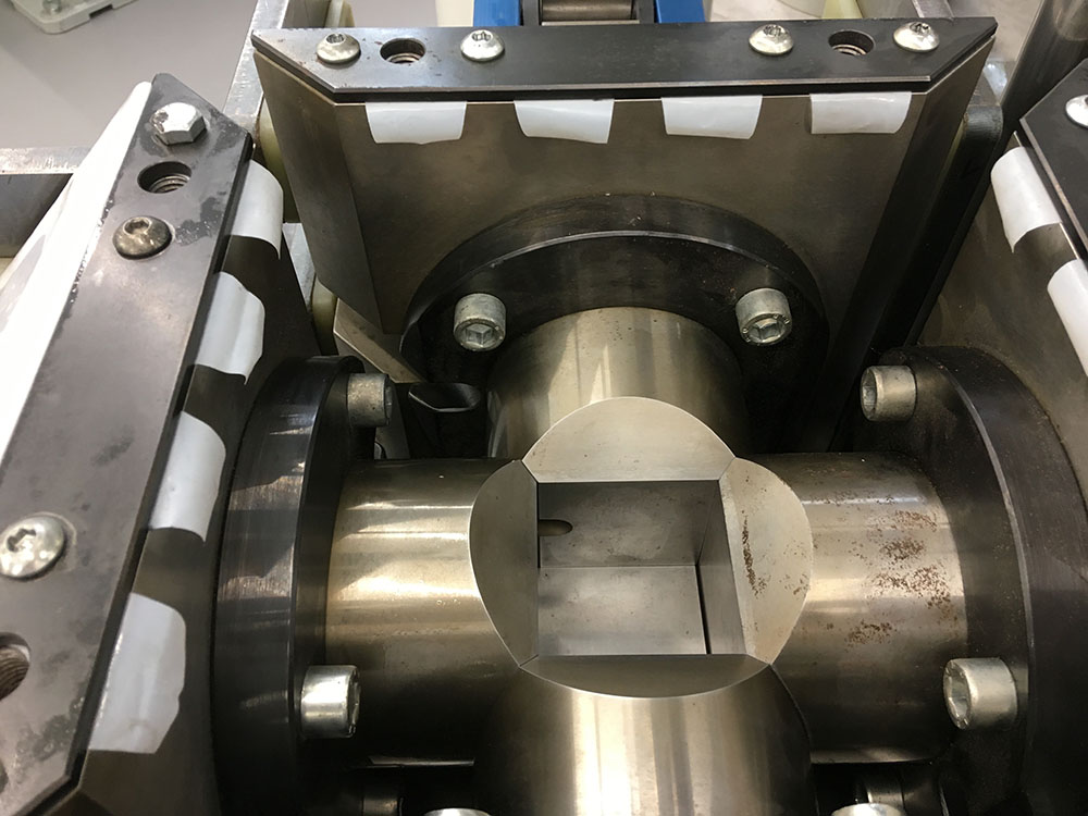
Cubic anvils MAX2003.
Equipment for high-pressure experimentation
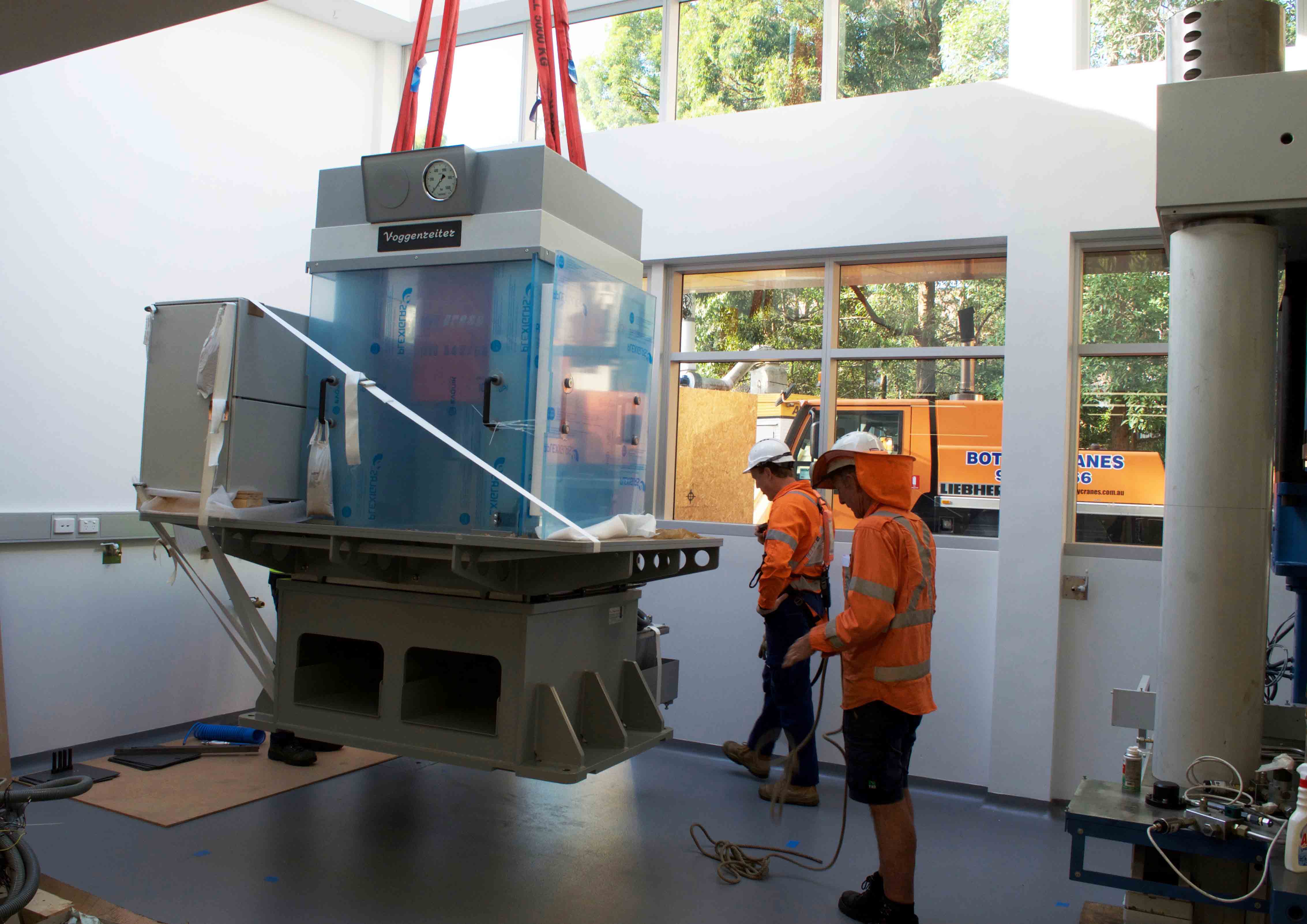
The experimental laboratory was closed in February 2018 for renovation and expansion. In April 2019, two multi-anvil presses were lowered into position through a removable roof. The larger MAX2003 is a cubic anvil set-up (pictured below), and is complemented by a Walker module in the smaller 1000 ton-press (pictured below). The laboratory is expected to reopen mid 2019.
The extended laboratory now includes three multi-anvil apparatuses, one diamond-anvil cell and two piston-cylinder apparatuses. Two additional rapid-quench piston-cylinder apparatuses are due for delivery in August 2019. There are also two Griggs apparatuses, and one-atmosphere quench furnaces.
Experimental projects include partial melting of peridotites in controlled volatile-present conditions at mantle pressures, electrical conductivity measurements on mantle minerals and rocks, reaction experiments that juxtapose subducted sedimentary materials with mantle peridotite to study melting behaviour, and the stability of nitrogen-bearing phases at high pressures, including the partitioning behaviour of nitrogen between minerals and melts.
PROGRESS IN 2018:
1. Facility for Integrated Microanalysis
a. Electron Microprobe: Dr Timothy Murphy, appointed in 2017, oversees the electron microprobe. The SX100 was fully refurbished by Cameca engineers (thanks to Nicolas Boutron and Pierre-Yves Corre) in November 2017 and now performs extremely well on a day to day basis. High-quality mapping of amazing microstructures in corundum from Israel (Griffin et al., CCFS publication #830) were published in the CAMECA user guide and marketing booklet. The EPMA is being updated with “Probe” software developed by John Donovan.
CCFS and AuScope have provided significant funding support and scientific expertise to purchase a Scanning X-ray spectrometer to enable fast scanning and mapping of thin sections and blocks, thus providing a wider and more complete spatial framework for in situ analysis. The acquisition and running of this instrument is a joint venture with Professor Damien Gore (Dept. Environmental Sciences). The versatility of this instrument has attracted significant interest from most faculties across Macquarie University, including Arts, and is heavily used by MRes and PhD students. Dr Timothy Murphy is leading a group developing new approaches with this instrument.
b. Laser-ablation ICPMS microprobe (LAM): Dr Yi-Jen Lai manages the extensive LA-ICP-MS and MC-ICP-MS instrument park available at Macquarie. CCFS Research Associate Yoann Gréau provided invaluable technical help and expertise, with CCFS technical and research staff Romain Tilhac and Hadrien Henry providing generous assistance for users.
The Photon Machines Excite/G2 laser system and Agilent 7700 ICPMS are used for in situ trace element analyses and U-Pb geochronology. The facility was used by 10 Macquarie PhD thesis projects, 6 international visitors, 4 Masters Research students and several in-house funded research projects and industry collaborations. Projects included the analysis of minerals from mantle-derived peridotites, pyroxenites and chromitites, meteorites, unusual types of ultra-reduced phases from volcanic sources and ultra-high pressure terranes, high-grade metamorphic rocks and biominerals.
With the addition of trace gases such as N2 and H2 in the ablation gas, Olivier Alard and collaborators have obtained a significant increase in terms of sensitivity (counts per ppb multiplied by 2) and a noticeable decrease in detection limit. This breakthrough allows researchers to investigate: (i) olivine trace element abundances (i.e. higher sensitivity means complete REE patterns can now be obtained), (ii) ultratrace element concentrations and distributions between silicates, sulfides and oxides of rarely investigated elements such as metalloids form the d- and p-blocks elements (e.g. Sn, Sb, Cd , Mo, W …). This technique is now being applied by Marina Vetter (Ph.D.) S. Foley and S. Demouchy (CNRS, Géoscience Montpellier) who visited this year.
The new Q3-ICP-MS (Agilent 8900) was installed in December 2017 and is co-located with the upgraded Nu-Plasma HR. The development of in situ Rb-Sr analysis is well underway. Preliminary results will be presented by Lauren Gorojovsky and Olivier Alard at the Goldschmidt conference in August 2019 (Barcelona, Spain). In-house reference materials have been characterised to extend the range of material (matrix) analysed. The team lead by Olivier Alard is also working on other developments for the precise (interference-free) measurement of chalcophile and siderophile elements for precise S-Se and Te analyses by LA-ICP-MS in submarine glasses (L. Gorojovsky MRes).
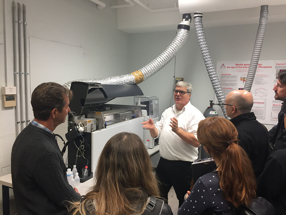
Fred Fryer (Agilent) demonstrating the ins and outs of the new Agilent Triple Quad ICPMS to the 2018 Geoanalysis delegates.
c. MC-ICPMS:
A Nu Plasma II MC-ICPMS was installed in June 2015 and followed the decommissioning of the Nu Plasma 005 after 16 years of service. Although the Nu Plasma II represents a significant advance in its electronics and engineering, much of the fundamental design is adapted from the Nu Plasma I. This enabled a relatively seamless transition of existing methods developed over the past 15 years on the Nu Plasma I. The combination of the expanded collector array (16 Faraday cups and 5 ion counters) and enhanced sensitivity compared to the first-generation Nu Plasma instruments has enabled the refinement of several in situ techniques pioneered at GEMOC, Macquarie.
Montgarri Castillo-Oliver and Yoann Gréau have refined the measurement of in situ Sr isotopes in carbonate and clinopyroxene by LA-MC-ICP-MS. New results are under review.
The in situ measurement of U-Pb isotopes in zircon using the combination of the femtosecond laser system and the Nu Plasma II was a world first, with preliminary results reported at the Goldschmidt Conference in Prague, August 2015 (N.J. Pearson, W.J. Powell, Y. Gréau, R.C. Murphy, J.L. Payne, E. Belousova, W.L. Griffin and S. Y. O’Reilly 2015. U-Pb geochronology of zircon by femtosecond laser ablation, Goldschmidt Abstracts, 2015, 2437). The development of standard operating procedures for in situ U-Pb, Re-Os and Rb-Sr isotope measurements is on-going. At the time of the installation of the new Nu Plasma II, the Nu Plasma HR 034 underwent an upgrade with an enhanced interface.
The upgrade increased sensitivity between 1.5 and 2 times, and this contributed to an overall improvement in signal stability, as well as in the precision of single measurements and long-term reproducibility. In 2015 a third Photon Machines excimer laser microprobe was installed and co-located with the Nu Plasma HR 034. After successful installation and commissioning had been achieved and the first experiments with the Femtosecond laser microprobe were completed (and presented at the 2015 Goldschmidt conference), key staff to develop the integrated system and applications were no longer available in 2016.
In addition, CCFS has funded a technology development program employing a Research Associate with a high level of instrument expertise (Dr Yoann Gréau) who, with Dr Olivier Alard (Future Fellow who brought the relevant expertise), has been recently making good progress with the envisaged developments. A novel split-stream approach has been established, involving the simultaneous measurement of Re-Os isotopes on the Nu plasma II and siderophile and chalcophile trace elements on the Agillent 7700. Preliminary results for this world first will be presented at the Goldschnidt conference in Barcelona (see Research highlight In situ laser ablation split-stream (LASS-) MC-ICP-MS for simultaneous determination of Re-Os isotopes and siderophile-chalcophile elements in sulfides: Ablating away a Cornelian dilemma). The technique will be used by several of our Chinese, Japanese and European collaborators visiting the laboratories in the first half of 2019. Planned applications are (i) combined U-Pb and Lu-Hf characterisation of zircons and (ii) simultaneous measurements of Sr isotopes and trace elements in silicates and carbonates. New technique strategies involving splitting with the Q3-ICP-MS are also being investigated.
CCFS/GEMOC remains one of the few facilities with the capability to perform in situ Re-Os dating of single grains of Fe-Ni sulfides and alloys in mantle-derived rocks. CCFS is translating the full methodology to the Taiwan Academia Sinica laboratories under the auspices of Dr Kuo-Lung Wang, a former GEMOC Research Fellow and CCFS Associate Researcher, to ensure preservation of the associated intellectual property and approach. CCFS Future Fellow Olivier Alard is undertaking studies of mantle sulfides worldwide, integrating in situ Re-Os and S isotopes obtained using the LA-MC-ICP-MS (Macquarie University) and ion probe (CAMECA 1280, CMCA Perth), respectively, in collaboration with CCFS Research Associate Laure Martin. This activity was also made possible by the expertise of CCFS Research Associate Yoann Gréau.
The LAM MC-ICPMS is the vehicle to deliver in situ high-precision ratio measurements including the analysis of Lu-Hf isotopes in zircon as a major part of TerraneChron® (see here.). TerraneChron® applications continued and have been recently up-scaled with the involvement of Dr Romain Tilhac, Nora Liptai and Hadrien Henry to meet the increasing demand for this powerful tool for understanding the evolution of Earth’s crust, for isotopic mapping and paleogeophysics, and geochemical remote sensing for the exploration industry.
d. Laboratory development: The clean-room facility established in 2005 continued to be used primarily for isotope separations for analysis on the Triton TIMS and the Nu Plasma MC-ICPMS. Routine procedures continued for Rb-Sr, Nd-Sm, Lu- Hf and Pb isotopes, as well as U-series methods (U, Th and Ra). Isotope dilution routines are being implemented by Peter Weiland and will soon be available.
e. Software:
GLITTER (GEMOC Laser ICPMS Total Trace Element Reduction) software is our online interactive program for quantitative trace element and isotopic analysis and features dynamically linked graphics and analysis tables. This package provides real-time interactive data reduction for LAM-ICPMS analysis, allowing inspection and evaluation of each result before the next analysis spot is chosen. GLITTER’s capabilities include the on-line reduction of U-Pb data. Sales of GLITTER are handled by AccessMQ and GEMOC provides customer service and technical backup. During 2018 a further 4 full licences of GLITTER were sold, bringing the total number in use to more than 300 worldwide, predominantly in Earth sciences applications but with growing usage in forensics and materials science.Dr Will Powell continued in his role in GLITTER technical support and software development through 2018 on a consultancy basis, following his resignation and relocation to Rio Tinto (Melbourne) in early 2016. The current GLITTER release is version 4.4.5 and is currently available without charge to existing customers.
2. X-Ray Fluorescence Analysis
2.1. In November 2012, a PANalytical Axios 1 kW X-ray Fluorescence (XRF) Spectrometer was installed and is used routinely to measure whole-rock major element compositions on fused glass discs and trace-element concentrations on pressed- powder pellets. In 2013 the sample preparation equipment was upgraded and included a new furnace to make high-quality cast glass beads. The major element calibration was modified in 2015 to extend the spectrum of rock types that could be analysed to include Fe-rich samples such as iron ores and laterites. This year the PANalytical was refurbished to maintain high accuracy and precision. Recent round robin tests (GeoPT) show that the PANalytical Axios is performing very well.
2.2. The high performance CHNS elemental analyser from Elementar (Vario El Cube) fitted with an extra IR-detector for low-level sulfur analysis is now in operation and is providing high quality S analyses for projects involving Re-Os isotopic analysis but also the distribution and abundance of volatile elements in the Earth’s mantle (PhD students Alimulati Ananuer, Michael Förster). A large suite of reference materials (n≈30), with variable matrix and composition, has been measured and the results presented at the Geoanalysis Conference (held at Macquarie University in July 2018). This facility enables us to better estimate whole-rock sulfur contents and thus sulfide abundance crucial to interpreting whole-rock and in situ Re-Os isotopic analysis. The reproducibility and low detection limit of the El Cube Elementar analyser yields remarkably accurate measurement of C, H and N at low levels for relatively small samples (i.e. ≈20 mg). See Research highlight Volatile elements and metasomatism: how to move things around in the lithosphere)
3. Whole-rock solution analysis
An Agilent 7500cs ICPMS produces trace-element analyses of dissolved rock samples for the projects of CCFS/GEMOC researchers and students and external users, supplementing the data from the XRF. We are testing the performance of the Nu Attom for ultratrace element measurements. The ICPMS dedicated to solution analysis is also used to support the development of ‘non-traditional’ stable isotopes with the refinement of separation techniques and analytical protocols (see 1. d).
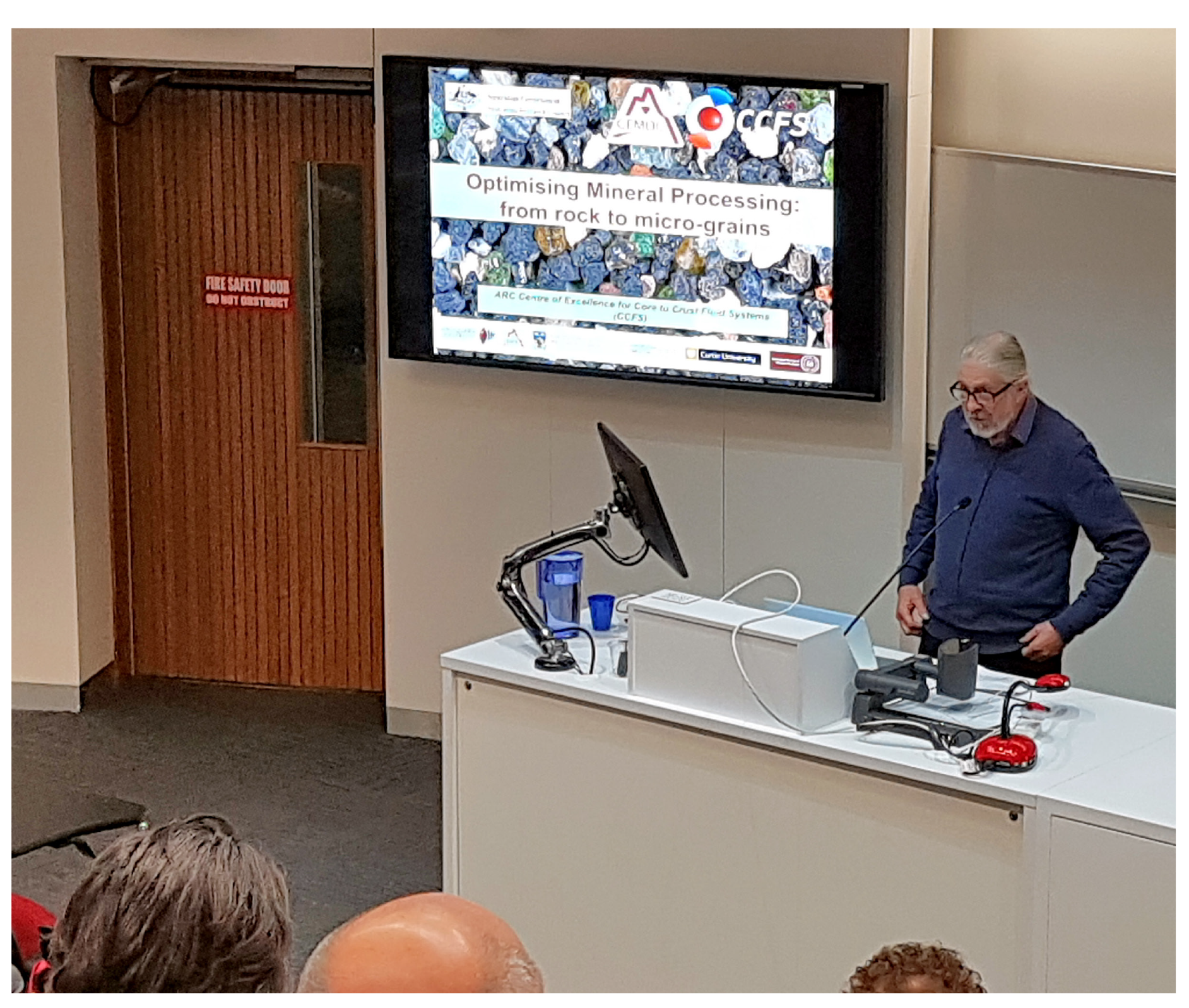
Steve Craven discussing the applications of the selFrag at the 2018 Geoanalysis Pre Conference workshop.
4. selFrag - a new approach to sample preparation
GEMOC’s selFrag instrument was installed in May 2010 and was the first unit in Australia. This instrument uses high-powered electrical pulses to disaggregate rocks and other materials along the grain boundaries. It removes the need to crush rocks for mineral separation and provides a higher proportion of unbroken grains of trace minerals such as zircon. Since its installation selFrag has been used for a range of applications including zircon separation, the analysis of grain size and shape in complex rocks, and the liberation of trace minerals from a range of mantle-derived and crustal rocks.
At the end of 2018 Dr Steve Craven retired. Steve helped building up the selFrag and was of immense value to numerous users in supporting them to get the best possible results for their sample preparation and consecutive analytical sessions at MQGA. This year the selFrag lab is supported by Miss Lauren Robinson, supported by Dr Timothy Murphy and Dr Sean Murray. Dr Steve Craven drops by from time to time to keep his guidance accessible.
5. Spectroscopy
The spectroscopy infrastructure includes an FTIR microscope (ThermoFisher iN10 FTIR microscope; 2008). The FTIR is used to measure H abundance in a range of nominally anhydrous minerals (e.g. olivine, pyroxene, garnet) and H and N contents in diamond. In developing the spectroscopy capability, an emphasis has been placed on hyperspectral mapping to produce integrated datasets and multi-layered information in a spatial context. A Horiba H-CLUE CL monochromator was installed on the Zeiss EVO SEM in January 2016. The monochromator system provides spatially resolved quantitative cathodoluminescence spectra, which allow identification of emitters (e.g. REE in zircons), crystal lattice vacancies (e.g. in diamond) and crystallographic information on how specific elements are incorporated in the mineral crystal lattices (e.g. Mn in aragonite). The new instrumentation is acquiring a growing group of users and is currently part of projects in biomineralisation (HDR student Laura Otter/Prof Dorrit Jacob), diamond growth (Professor Dorrit Jacob) and zircon characterisation (Honorary Associate Dr Christoph Lenz/Dr Elena Belousova).
6. Raman spectrometry
A confocal laser Raman microscope (co-funded by the Macquarie University Strategic Infrastructure Scheme (MQSIS), 2014 and Future Fellowship funding to Professor Dorrit Jacob) delivers information for non-destructive phase-identification and -characterisation at one micrometre spatial resolution. The Raman spectrometer continues to serve the CCFS, the Department and the Faculty. In 2018 the system’s capabilities were extended with the purchase of two new laser wavelengths (785 and 432 nm; MQSIS 2017). The instrument continued to grow its user base across the Faculty of Science and Engineering at Macquarie University with users from Chemistry, Physics, Biology, Environmental Sciences as well as users from the Faculty of Arts, Department of Ancient History and the Museum of Ancient Cultures.
In 2019 we plan to extend the applications of Raman Spectrometry towards other areas of scientific research.
These include:
- Earth and Planetary Science, analysis of sulfate speciation in glasses (Dr Oliver Alard and Lauren Gorojovsky)
- Forensics applications, namely ink characterisation on Egyptian papyrus (with Prof Damian Gore and Assoc Prof Malcolm Choat)
- Chemistry, surface enhanced Raman spectroscopy (SERS) of nano particle interactions in serum (Dr Alfonso Garcia-Bennett and Inga Kuschnerus)
- Molecular Science, plasmonic nanostructures for surface-enhanced Raman spectroscopy
- Physics, analysis and mapping of element distribution in wave guides (Prof Simon Gross, Dr Toney Fernandez and Thomas Gretzinger)
- Archaeology, oxide and corrosion analysis of ancient lead scrolls (Prof Simon Clark and Carla Raymond)
- Archaeology, identification of pigments used in Egyptian Mummy Carapace (Dr Karin Sowada and Dr Ronika Powers)
- Archaeology, pigment analysis of Amarna Blue used in Egyptian pottery (Prof Martin Bommas and Penelope Edwell)
7. Computer cluster
Computational geodynamics has been supported throughout this project through a number of in-house machines (Enki and Toto), as well as a Macquarie partnership with NCI, that has enabled large project-based allocations on the national machines. The former resources have enabled the development and testing of in-house computational tools, including Aspect modules (led by Craig O’Neill and former postdoc Siqi Zhang) to model crustal production, impact melting and magmatic melt emplacement, and also Litmod in modelling crustal and lithospheric structure. Our access to the large-scale facilities has enabled production-level simulations and has supported > 5 PhD projects, postdocs and numerous Masters projects.
CMCA TECHNOLOGY DEVELOPMENT AND INSTRUMENTATION
The University of Western Australia’s Centre for Microscopy, Characterisation and Analysis (CMCA) is a $50 M core facility providing analytical solutions across a diverse array of scientific research. The world-class facilities and associated technical and academic expertise are the focus of micro-analytical and characterisation activities within Western Australia, while strong links and collaborations have earned the CMCA an excellent national and international reputation. The CMCA incorporates the Western Australian Centre for Microscopy, and is a node of the NCRIS Characterisation capabilities, the National Imaging Facility (NIF) and the Australian Microscopy and Microanalysis Research Facility (AMMRF). It is also associated with the NCRIS funded Australian National Fabrication Facility (ANFF), and AuScope, which have made a substantial contribution to facilities run by CMCA.
CMCA capabilities:
- Secondary Ion Mass Spectrometry (CAMECA IMS 1280 and CAMECA NanoSIMS 50 and NanoSIMS 50L)
- Electron probe microanalysis (JEOL JXA 8530F)
- Focused ion beam (FEI Helios)
- Transmission electron microscopy (FEI Titan, JEOL 2100)
- Scanning electron microscopy (FEI Verios XHR, Zeiss 1555, Tescan Vega3)
- X-ray powder diffraction (Panalytical Empyrean)
- X-ray micro-CT (Xradia)
- Confocal Raman imaging with AFM (WiTec Alpha 300RA+)
- NMR spectroscopy (2 Bruker Avance and 2 Varian spectrometers)
- X-ray crystallography (Oxford Diffraction)
- GC and HPLC mass spectrometry
- Bioimaging, flow cytometry, cell sorting, and laser micro-dissection
- Optical and confocal microscopy
- Biological sample cryo-preparation and ultramicrotomy
THE AMMRF FLAGSHIP ION PROBE FACILITY
The CAMECA IMS1280 and NanoSIMS 50 are flagship instruments of the AMMRF. The AMMRF Flagship Ion Probe Facility offers state- of-the-art secondary ion mass spectrometry (SIMS) capabilities to the Australian and international research communities, allowing in situ, high-precision isotopic and elemental analyses, and secondary ion imaging on a wide range of samples.
The IMS1280 large-geometry ion probe, installed in 2009, was co-funded by the University, the State Government of Western Australia, and the Federal Government’s Department of Innovation, Industry, Science and Research (DIISR) under the “Characterisation” (AMMRF) and “Structure and Evolution of the Australian Continent” (AuScope) capabilities of the National Collaborative Research Infrastructure Strategy (NCRIS). The NanoSIMS 50, installed in 2003, was funded through the Federal Government’s NCRIS-precursor, the Major National Research Facility scheme (NANO-MNRF). UWA’s Ion Probe Facility can currently lay claim to being the best-equipped SIMS lab in the world, as no other facility has two NanoSIMS alongside an IMS1280.
The Ion Probe Facility is a key characterisation component within the ARC Centre of Excellence for Core to Crust Fluid Systems. To ensure the highest levels of quality and throughput, CCFS has provided funding for a Research Associate position within the Ion Probe Facility, to facilitate direct scientific and technical interaction for all CCFS users and projects.
Olivier Alard visiting CMCA to use the CAMECA SIMS 1280.
PROGRESS IN 2018:
The Ion Probe Facility has continued to contribute to various projects in the context of CCFS. Both 1280 and nanoSIMS laboratories contributed to ~40 individual projects in Earth Sciences, originating from CCFS partners, other Australian research institutes and overseas. 34 journal articles were published in 2018 featuring data acquired using the IMS1280, the NanoSIMS 50 and 50L at UWA, of which 10 were directly related to CCFS projects.
CMCA was successful in winning an ARC LIEF grant for a new EPMA to support the characterisation of minerals and materials for researchers in Western Australia. It is anticipated that the new instrument will be installed in early 2019.
For further information on CMCA facilities please consult
http://www.cmca.uwa.edu.au/
JOHN DE LAETER CENTRE
The John de Laeter Centre (JdLC) is based at Curtin University and forms one of the university’s core research centres. The centre houses advanced instrumentation for high-quality chemical, mineralogical and microstructural analysis, and high-resolution imaging. It hosts over $28M in infrastructure supporting research in: geosciences (geochronology, thermochronology and isotope studies); environmental science; isotope metrology; forensic science; economic geology (minerals and petroleum); marine science; and nuclear science.
The facility website provides detailed information on the new facilities, instrumentation and research staff (http://www.jdlc.edu.au).
The components of the JDLC are organised into fourteen major facilities including:
(GAP) Geoscience Atom Probe Facility:
GAP is a node of the Advanced Resources Characterisation Facility (ARCF) funded by a $12,400,000 Science and Industry Endowment Fund grant to Curtin, UWA and CSIRO. The GAP hosts a Cameca LEAP 4000X HR microscope capable of carrying out atom probe tomography (APT), a recent development in the geosciences, that provides high spatial resolution with time- of-flight mass spectrometry to provide 3-dimensional chemical information at the atomic scale. More commonly used to study semiconductors and metal alloys, the GAP is the first atom probe facility in the world to be dedicated to the study of geological materials (http://www.geoscienceatomprobe.org). The ARCF also commissioned a Tescan Lyra focused ion beam scanning electron microscope (FIB-SEM), with a Ga+ gun capable of micro- milling out a 100 nm wide needle of a mineral sample prior to APT analysis. The Lyra system is a highly advanced platform for 2D and 3D microanalysis with time of flight mass spectrometry (TOF-SIMS) and electron back scattered diffraction (EBSD) detectors. By correlating the analytical outputs of both the LEAP and the Lyra instruments, the ARCF provides an unprecedented capability of characterising highly complex materials at the nanoscale.
(DMH) Digital Mineralogy Hub Facility:
The Facility hosts a Tescan Integrated Mineral Analyzer (TIMA GM) - a fully automated, high throughput, analytical Field Emission Gun Scanning Electron Microscope (FEGSEM) for automated analysis of sample composition. TIMA measures mineral abundance, liberation properties, mineral association and grain size automatically on multiple samples of grain mounts, thin sections or polished sections. Applications include ore characterisation, process optimisation, remediation and the search for precious metals and strategic elements. The facility is being used by a broad spectrum of researchers: geologists and archaeologists are using the facility in petrological characterisation, sample classification and lithofacies studies; while geochemists and geochronologists are using the mineral classification outputs as targeting maps for further ion, electron or laser microprobe analysis.(CEG) Curtin Experimental Geochemistry Facility:
CEG provides facility for experimental petrology, geochemistry and hydrogeochemistry at pressures and temperatures that range from those at the Earth’s surface to those at the base of the Earth’s crust. The Facility contains:
- 2 x 150 ton end loaded piston cylinder presses
- Coretest hydrothermal apparatus
- Assorted furnaces to 1400 degrees C
- Assorted titanium and Teflon-lined bombs
(GHF) GeoHistory Facility:
The GHF houses state-of-the-art laser ablation inductively coupled plasma mass spectrometry (LA-ICPMS) equipment, in addition to a low temperature thermochronology laboratory. The LA-ICPMS comprises a Resonetics S-155-LR 193nm excimer laser ablation system coupled to an Agilent 7700x quadrupole ICPMS. The Excimer laser is also coupled to a RESOchron helium analysis line for in situ (U-Th-Sm)/He, U-Pb and trace element analysis of single crystals. The facility also has a separate Alphachron helium line with a diode laser and furnace in order to facilitate conventional (U-Th)/He dating on single mineral crystals and larger samples. A Nu Plasma II multi-collector was integrated into the facility to facilitate split stream analysis.
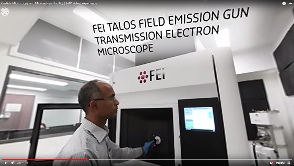
Take a 360° virtual tour of Curtin’s Microscopy and Microanalysis Facility here.
(MMF) Microscopy and Microanalysis Facility:
The MMF houses a broad range of advanced microanalysis instrumentation providing high quality chemical, mineralogical and microstructural information, and high resolution images for research and technical publications. The facility staff have expertise in Materials and Earth Science research which is used to support both academic research and applied projects for the Western Australian minerals and energy sector. Techniques and instrumentation available include:
- High resolution imaging is available through a FEI Talos F200X transmission electron microscope (TEM), which was commissioned in early 2017 to complement ongoing research at the nanoscale. The system combines high resolution S/TEM and TEM imaging with EDS and 3D chemical characterisation. The instrument is capable of elemental and microstructural analysis at extremely high magnifications.
- Spatially resolved elemental analysis (EDS) and phase and orientation analysis (EBSD) on a Tescan MIRA3 platform - a variable pressure field emission scanning electron microscope (VP-FESEM) that features sensitive EDS and Oxford Symmetry EBSD detectors and integrated software for high quality microstructural analysis of crystalline samples. In 2019 this instrument will be complemented by a new state-of-the-art FESEM with a second Symmetry EBSD system, which will allow EBSD analysis at up to 3000 Hz.
- Quantitative mineral analysis (Q-XRD) - The D8A is an X-ray Diffractometer (XRD) with a copper X-ray source and an automated 45 position sample changer. It features a LynxEye position sensitive detector that is 200 times faster than a conventional scintillator detector, allowing collection of superior data in a short time-frame.
- Ion beam sample manipulation including TEM & TKD lamella preparation (FIB) - The NEON is a dual beam focused ion beam scanning electron microscope (FIB-SEM) equipped with a field emission gun and a liquid metal Ga+ ion source. This instrument combines high resolution imaging with precision ion beam ablation of focused regions, allowing for site specific analysis of the surface and subsurface of samples in 2D or 3D.
The MMF also houses a suite of equipment that includes light microscopy, vacuum mount impregnation, manual and automated polishers, mills and coaters that are used to prepare samples for electron microscopy and X-ray diffraction.
(SAXS) Small Angle X-Ray Scattering Facility:
Small angle X-ray scattering can be used to characterise the size, shape and distribution of objects between 1 and 100 nm. In 2016, LIEF funding was used to upgrade the instrumentation in the facility. The WA SAXS facility houses a Bruker NANOSTAR SAXS instrument comprising an Excillum MetalJet high-intensity X-ray source, in-vacuum specimen chamber, and a two- dimensional photon counting detector, capable of covering a q-range of 0.008 – 1.25 Å-1.

(SHRIMP) Sensitive High Resolution Ion Micro Probe Facility:
The SHRIMP are large mass spectrometers that allow in situ isotopic and trace element micro-analysis of complexly zoned minerals in grain mounts and thin section plugs, with a spatial resolution of 5-20 microns. The facility at Curtin has two automated SHRIMP II ion microprobes capable of 24- hour operation, together with a preparation laboratory that was remodelled in 2014. The main application of the SHRIMP instruments at Curtin is for U-Th-Pb geochronology of zircon and other U-bearing minerals, including monazite, xenotime, titanite, allanite, rutile, apatite, baddeleyite, cassiterite, perovskite and uraninite where multiple growth zones commonly require analyses with high spatial resolution. SHRIMP II is fitted with a Cs source, electron gun and 5 channel M/C.
(SMS) SelFrag & Mineral Separation Facility:
A SelFrag facility, supported by an ARC LIEF grant, has been installed within the Department of Applied Geology at Curtin University. High voltage electrodynamic disaggregation of materials in gram- to kilo-scale batches, along with downstream mineral separation processing, to deliver mineral concentrates, separates, mounts for SHRIMP and LA-ICPMS analysis. The facility provides electric pulse disaggregation for mineral separation, which allows mineral grains to be separated from rock samples without the damage associated with standard crushing techniques.
(TIMS) Thermal Ionisation Mass Spectrometry Facility:
The TIMS Facility provides highly accurate and precise measurements of the isotopic composition of elements using TIMS TritonTM instrument, including sample preparation in a clean, contamination-free environment. The Triton is equipped with a 21-sample turret and 9 faraday cups, enabling a precision of 0.001% on isotopic ratios. As well as geological applications within the broad field of isotope geochemistry (Re/Os, U/ Pb, Pb/Pb, Sm/Nd, Rb/Sr) the TIMS instruments can be applied to a variety of isotope fingerprinting, such as forensics and the environmental impact of human activities. The TIMS instruments are also used for the calibration of isotopic standards and the calculation of isotopic abundances and atomic weights. The facility has recently installed a Thermo Scientific Triton™ mass spectrometer, facilitating a new range of geochemical, geological and environmental research applications.
(TRACE) TRACE Research Advanced Clean Environment Facility:
This consists of a ~400 m2 class 1000 containment space housing four class 10 ultra-clean laboratories, a class 10 reagent preparation laboratory and a −18 °C class 10 cold clean laboratory, located at Curtin University. The extremely low ultimate particle counts are achieved with successive ‘spaces within spaces’ and HEPA filtration at each stage.
(WAAIF) Western Australian Argon Isotope Facility:
This is located at Curtin and is equipped with A MAP215-50 mass spectrometer and an Argus VI Multi-Collector Noble Gas Mass Spectrometer with a low-blank automated extraction system coupled with a New Wave Nd-YAG dual IR (1064 nm) and UV (216 nm) lasers, electromultiplier detectors and Niers sources. The ultra-violet laser is capable of high-resolution (up to 10 µm beam size) ablation of any mineral, allowing detailed analysis of individual mineral grains. The 40Ar/39Ar method is used to date a myriad of geological events such as volcanism, tectonic plate movements, mountain building rates, sediment formation, weathering and erosion, hydrothermal fluid movements, and alteration and diagenesis of minerals.
(WA-OIG) WA Organic and Isotope Geochemistry Facility:
WA-OIG is an internationally-recognised group contributing to world-class research in the fields of organic and stable isotope geochemistry, paleogenomics and geomicrobiology. Available techniques are listed here: http://jdlc.edu.au/wa-organic-and- isotope-geochemistry-facility-wa-oig/.
For further information on JDLC facilities please consult
http://www.jdlc.edu.au
WESTERN AUSTRALIA PALEOMAGNETIC AND ROCK-MAGNETIC FACILITY
The Western Australia Paleomagnetic and Rock-magnetic Facility is a national research infrastructure supported by the Australian Research Council and collaborating institutions including Curtin University, the University of Western Australia (UWA), the Australian National University, Macquarie University and University of Queensland. The facility was established at UWA in 1990 by CCFS CI Z.X. Li, and has been progressively upgraded over the years. The facility is now completely housed in purpose-built laboratories on Curtin University’s Bentley campus.
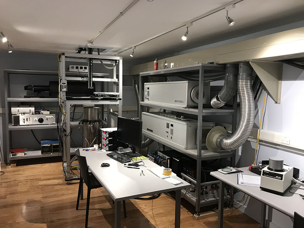
Inside the Shielded Room.
A significant component of the facility is the magnetically shielded room (constructed in mid-2015 by Dr Gary Scott’s team) which provides a 20m2 laboratory space with ambient magnetic fields less than 0.5% of the local geomagnetic field. Within this shielded room are: a 2G 755 superconducting rock magnetometer with a vertical Model 855 automated sample handler (the RAPID system), an AGICO JR-6A spinner magnetometer, and ASC TD-48SC and MAGNETIC MEASUREMENTS thermal demagnetisers. An earlier model 2G 755 cryogenic magnetometer, which underwent repair and upgrade during 2017-18, will be installed within the shielded room during the first half of 2019.
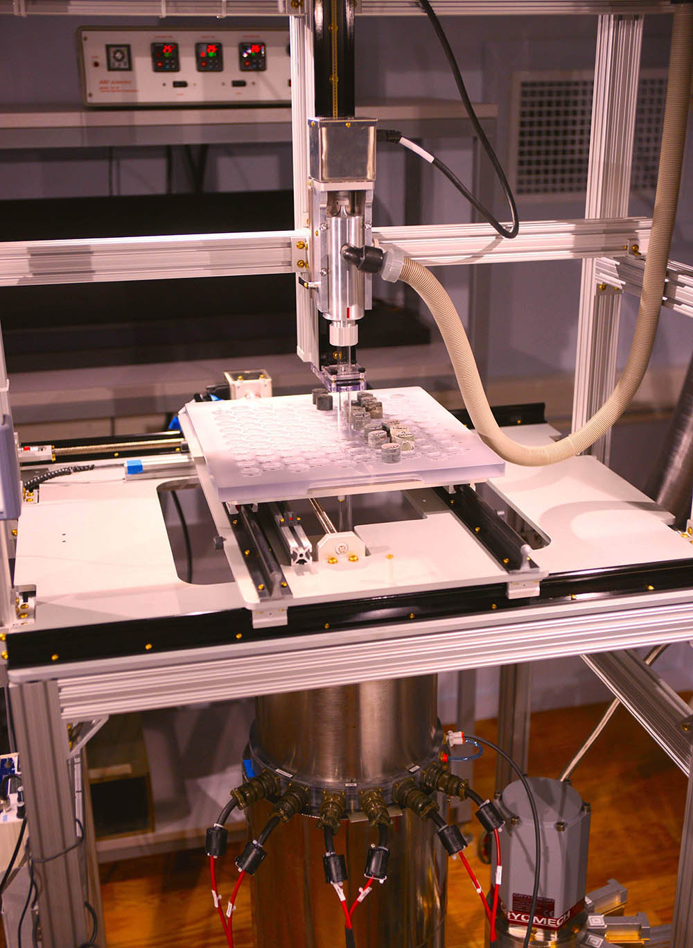
RAPID sample stage.
Other apparatus are housed in the renovated laboratory spaces surrounding the shielded room and include: a MAGNETIC MEASUREMENTS MMPM5 pulse magnetiser, an AGICO MFK-1FA Kappabridge, and the Petersen Instruments Variable Field Translation Balance (VFTB). In mid-2018 both the Kappabridge and VFTB were upgraded to bring them up to the current state-of-the-art. A temperature-susceptibility (K-T) module was added to the Kappabridge and a full electronics upgrade was performed on the VFTB system, improving the sensitivity and response time, as well as providing additional functionality (First Order Reversal Curve measurement). An additional module has also been recently installed on the RAPID system to enable acquisition, and subsequent measurement, of Isothermal Remanent Magnetisation (IRM).
The recent purchases, upgrades and co-location of all instruments represent a major enhancement to the productivity and capabilities of the facility. Apparatus in the facility include:
- a 2G 755 superconducting rock magnetometer with a vertical Model 855 automated sample handler (the RAPID system) and other accessories (including; AF coils, susceptibility meter, ARM and IRM modules)
- an earlier model 2G 755 cryogenic magnetometer upgraded to a 4K DC SQUID system (plus a recent upgrade carried out by 2G enterprises, including the repair of the lightning-damaged cold head)
- an AGICO JR-6A spinner magnetometer
- 1x MMTD80, 2x MMTD18 and a TD-48-SC thermal demagnetiser
- a Petersen Instruments Variable Field Translation Balance (VFTB)
- an AGICO MFK-1FA Kappabridge with K-T capacity
- a MAGNETIC MEASUREMENTS MMPM5 pulse magnetiser
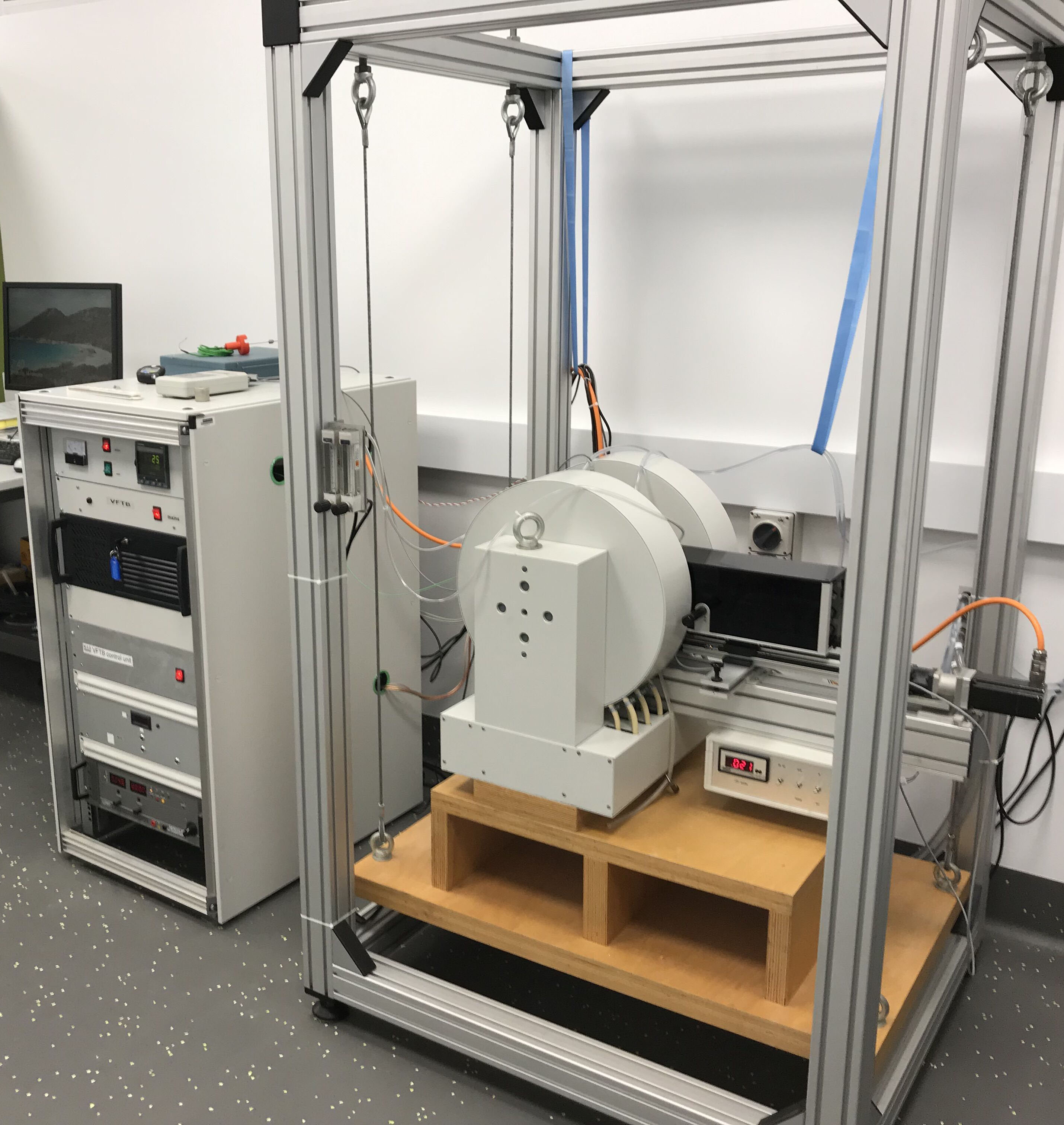
VFTB system.

The facility supports a wide range of research topics, including reconstruction of global paleogeography (the configuration and drifting history of continents) through Earth’s history, reconstructing the evolving geomagnetic field (e.g. paleointensity) through time, analyses of regional and local structures and tectonic histories, dating sedimentary rocks and thermal/chemical (e.g. mineralisation) events, studying past climate changes, and orienting rock cores from drill-holes.
A national workshop on paleomagnetism, rock magnetism and their applications to tectonics, paleoclimate research, and Earth resource exploration will be conducted in February 2020. It will include a tour of the facilities along with training on the operation of all instruments for potential users of the laboratory.